
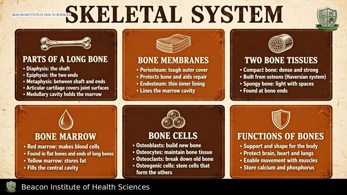
Reference-based BSN study notes for Anatomy and Physiology I.
Learning objectives and functions
Describe skeletal functions, classify bones, identify axial and appendicular divisions, explain long-bone structure, and outline ossification, growth, remodeling and fracture repair. Bones support the body, protect organs, provide levers for muscle movement, store calcium and phosphate, and house marrow. Red marrow supports blood-cell production; yellow marrow is predominantly fatty. Bone is living vascular connective tissue, not an inert frame.
Bone classification
Long bones, such as the femur and humerus, have a shaft and expanded ends. Short bones include many carpals and tarsals. Flat bones, including the sternum and many skull bones, protect organs and provide muscle attachment. Irregular bones include vertebrae. Sesamoid bones develop within tendons; the patella is the largest usual example. Shape classification does not depend simply on absolute size: phalanges are long bones despite being small.
Long-bone anatomy
The diaphysis is the shaft; epiphyses are the ends; metaphyses lie between them. In growing bones, epiphyseal plates contain cartilage supporting length growth. The adult epiphyseal line marks a closed plate. Articular cartilage covers joint surfaces and reduces friction. Periosteum surrounds most external bone surfaces except articular areas and supports nourishment, attachment and repair. Endosteum lines internal surfaces. The medullary cavity contains marrow. Nutrient vessels and nerves pass through foramina; a fracture can damage both skeletal and neighbouring neurovascular structures.
Compact and spongy bone
Compact bone forms a strong external cortex. Its osteons have concentric lamellae around central canals carrying vessels and nerves. Osteocytes occupy lacunae, connected through canaliculi; perforating canals connect vascular pathways. Spongy bone consists of trabeculae arranged according to loads, with marrow spaces between them. It is not simply weak bone or a collection of complete osteons. Collagen contributes tensile resilience, while mineral, chiefly hydroxyapatite, provides resistance to compression.
Osteoprogenitor cells give rise to bone-forming cells. Osteoblasts produce matrix; osteocytes maintain and sense their local environment; osteoclasts resorb bone. Remodeling balances formation and resorption throughout life. Mechanical loading, nutrition, age and hormones influence this balance. Reduced loading during prolonged immobility can contribute to bone loss.
Axial skeleton
The usual adult count is 206 bones, with variation due to fusion and accessory bones. The axial division has 80 bones: skull, auditory ossicles, hyoid, vertebral column and thoracic cage. The cranium protects the brain; facial bones form the face and support openings. The hyoid supports tongue-related structures without a usual direct articulation with another bone.
The vertebral column typically includes seven cervical, twelve thoracic and five lumbar vertebrae, plus the sacrum and coccyx formed by fusion. Intervertebral discs contribute cushioning and movement. The thoracic cage includes sternum and twelve rib pairs. Ribs 1-7 attach to the sternum through their own costal cartilage; 8-10 attach indirectly; 11-12 are floating ribs without an anterior sternal attachment. Learn regions and landmarks before memorising every process.
Appendicular skeleton
The appendicular division has 126 bones and includes girdles and limbs. Clavicles and scapulae form the pectoral girdles. Each upper limb has a humerus, radius, ulna, eight carpals, five metacarpals and fourteen phalanges. In anatomical position, the radius is lateral on the thumb side and the ulna medial.
Each hip bone combines ilium, ischium and pubis. The pelvic girdle transfers weight to the lower limbs. Each lower limb includes femur, patella, tibia, fibula, seven tarsals, five metatarsals and fourteen phalanges. The tibia is the major medial weight-bearing leg bone; the fibula lies laterally. The terms arm and leg anatomically refer to shoulder-to-elbow and knee-to-ankle regions, respectively, rather than the whole limb.
Formation, growth and calcium regulation
Intramembranous ossification forms bone within connective tissue, including many flat skull bones. Endochondral ossification replaces a cartilage model and produces most bones. Length growth occurs through organised cartilage growth and replacement at growth plates; appositional growth increases thickness. Closure limits further length growth, but remodeling continues.
Adequate protein, calcium, vitamin D and other nutrients support bone health. Vitamin D supports intestinal calcium absorption; parathyroid hormone participates in maintaining extracellular calcium through effects involving bone, kidney and vitamin D activation. Do not reduce calcium homeostasis to one hormone or assume supplementation is appropriate for every patient.
Repair and nursing relevance
Fracture healing commonly progresses through haematoma/inflammation, soft callus, hard callus and remodeling. Stability, vascular supply, nutrition and patient factors influence repair; timing varies substantially. A fracture is not healed simply because pain decreases. Nurses connect anatomy with immobilisation, safe movement and neurovascular observation, documenting pain, colour, temperature, sensation, movement and circulation according to local protocols. New numbness, worsening pain or impaired perfusion require prompt escalation.
Revise by labelling axial and appendicular bones, tracing a long bone from surface to marrow, and comparing osteoblasts with osteoclasts. Explain why growing and adult bones differ at the metaphysis, and why vascular injury can threaten repair. This unit provides normal anatomy; detailed fracture treatment and orthopaedic procedures require separate clinical guidance.