
Introduction to Cells, Tissues, and Membranes
This lesson provides foundational concepts in Anatomy and Physiology I, focusing on the essential building blocks of the human body. By studying these notes, BSN students will be able to identify a typical human cell and its organelles, explain selective membrane transport, outline cell division, compare the four tissue classes, and distinguish cell membranes from body membranes. Understanding the relationship between structure and functions like absorption, protection, movement, and tissue repair is critical for nursing practice.
Cell Organization and Organelles
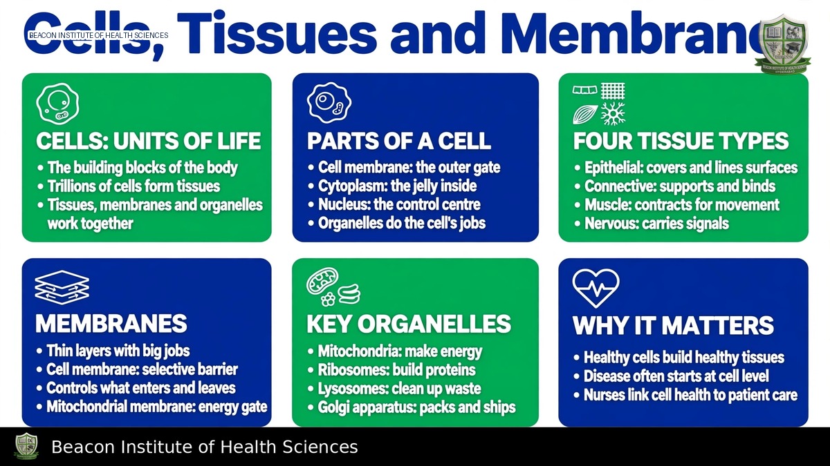
A typical human cell comprises a plasma membrane, cytoplasm, and a nucleus. The plasma membrane, a phospholipid bilayer, features hydrophilic surfaces and a hydrophobic interior. Proteins embedded within or associated with the membrane facilitate transport, act as receptors, function as enzymes, and provide attachment points. Cholesterol plays a role in influencing membrane behavior. The cytoplasm encompasses both the cytosol and various organelles. The nucleus houses DNA and is responsible for regulating gene expression, while the nucleolus is involved in assembling ribosomal components. It's important to note that not all mature human cells possess a nucleus; mature erythrocytes are a key exception.
Key Organelles and Their Functions:
- Ribosomes: Synthesize proteins.
- Rough Endoplasmic Reticulum (RER): Processes proteins destined for secretion or integration into membranes.
- Smooth Endoplasmic Reticulum (SER): Involved in lipid production, detoxification, and calcium handling in specialized cells.
- Golgi Apparatus: Modifies, sorts, and packages cellular products.
- Mitochondria: Support aerobic ATP production, supplying energy (ATP) for cellular work.
- Lysosomes: Digest cellular material.
- Peroxisomes: Participate in oxidative reactions.
- Cytoskeleton: Maintains cell shape, facilitates movement, and supports intracellular transport.
- Cilia: Move material along a surface.
- Microvilli: Increase surface area for absorption. Cilia and microvilli are distinct and not interchangeable structures.
Membrane Transport Mechanisms
Cell membranes regulate the passage of substances through various transport mechanisms.
Passive Membrane Transport
Passive transport mechanisms move substances down their concentration gradients without requiring direct ATP expenditure.
- Simple Diffusion: Movement of substances down their concentration gradients. Small lipid-soluble molecules and respiratory gases can readily cross the phospholipid bilayer via simple diffusion.
- Facilitated Diffusion: Utilizes channels or carriers to move substances down an electrochemical gradient. This is a protein-mediated process but is not necessarily active transport.
- Osmosis: The movement of water across a selectively permeable membrane, influenced by water chemical potential and effective solute differences.
- Filtration: Bulk movement driven by pressure differences, as observed in capillary and renal physiology.
Tonicity describes a solution's effect on cell volume and depends on effective, poorly penetrating solutes. Cells tend to swell in a hypotonic environment and shrink in a hypertonic environment, while isotonic conditions result in no sustained net volume change. While related, tonicity and total osmolality are not identical. As a critical nursing consideration, classroom comparisons of solutions do not authorize the choice or administration of intravenous fluids without prescribed, patient-specific guidance.
Active Transport and Vesicular Transport
Active transport mechanisms require energy to move substances, often against their concentration gradients.
- Primary Active Transport: Directly uses energy, typically ATP. The sodium-potassium ATPase is a prime example, moving three sodium ions out and two potassium ions in per ATP molecule, thereby maintaining vital ion gradients.
- Secondary Active Transport: Harnesses an existing ion gradient (established by primary active transport) to drive the uphill movement of another substance. Symport mechanisms move coupled substances in the same direction, whereas antiport mechanisms move them in opposite directions.
- Endocytosis: Involves the ingestion of material into vesicles, including phagocytosis of larger particles and receptor-mediated uptake.
- Exocytosis: Involves the release of vesicle contents, such as neurotransmitters or secreted proteins, from the cell.
Cell Cycle and Differentiation
The cell cycle encompasses phases of growth, maintenance, and division. During interphase, cells grow, maintain activity, and replicate DNA in the S phase. Mitosis is the process of separating replicated chromosomes through distinct stages (prophase, metaphase, anaphase, and telophase), followed by cytokinesis, typically yielding two daughter cells with the same chromosome complement. Meiosis is a specialized form of cell division that produces haploid gametes and contributes to genetic variation.
Differentiation is the process by which cells acquire specialized functions through regulated gene expression. The maintenance of tissues involves a balance of cell division, differentiation, and programmed cell death. Many mature specialized cells have limited regenerative capacity, meaning tissue healing rates can vary significantly.
Four Primary Tissue Classes
The human body is composed of four fundamental tissue types, each with unique structures and functions.
1. Epithelial Tissue
Epithelium covers body surfaces, lines cavities, and forms glands. Its cells are closely joined and rest on a basement membrane. Epithelial tissue is avascular, meaning it lacks its own blood vessels and receives nutrients from underlying connective tissue. Epithelium can be classified by the number of layers:
- Simple epithelium: One layer of cells.
- Stratified epithelium: Multiple layers of cells.
- Pseudostratified epithelium: Appears layered but all cells contact the basement membrane.
Cell shapes include squamous (flat), cuboidal (cube-shaped), and columnar (column-shaped). Examples include alveolar simple squamous epithelium, which facilitates exchange; intestinal simple columnar epithelium, which supports absorption; and epidermal stratified squamous epithelium, which provides protection.
2. Connective Tissue
Connective tissue consists of cells embedded within an extracellular matrix, which contains ground substance and fibers. The composition of this matrix determines the tissue's properties regarding support, flexibility, and transport. Examples include loose connective tissue, dense collagenous tissue, adipose tissue, cartilage, bone, and blood. Tendons and ligaments are classified as dense connective tissues. Cartilage generally heals slowly due to its lack of direct vascular supply.
3. Muscle Tissue
Muscle tissue is specialized for contraction.
- Skeletal muscle: Striated and typically under voluntary control.
- Cardiac muscle: Striated and involuntary, found only in the heart.
- Smooth muscle: Non-striated and involuntary, found in the walls of internal organs.
4. Nervous Tissue
Nervous tissue is composed of neurons, which transmit signals, and glia, which support the nervous system. It is important to remember that a single organ often contains several tissue types working together to perform its functions.
Body Membranes
Body membranes are thin sheets of tissue that cover surfaces, line cavities, and surround organs. It is crucial to distinguish a body's tissue membrane from a cell's plasma membrane.
- Cutaneous Membrane: This is the skin, providing external protection.
- Mucous Membranes: Line passages that communicate with the exterior, such as the digestive and respiratory tracts. Their epithelial structure varies depending on their specific function.
- Serous Membranes: Line closed ventral cavities and cover associated organs. Parietal layers line cavity walls, while visceral layers cover the organs themselves. Examples include the pleura (lungs), pericardium (heart), and peritoneum (abdominal organs).
- Synovial Membranes: Line the surfaces of appropriate joint capsules and produce components of synovial fluid. These are classified as connective tissue membranes, not true epithelial serosa.
Nursing Application and Revision Points
For BSN students and nursing educators, understanding these concepts is vital. Consider how diarrheal losses can significantly disturb extracellular conditions and impair cell function, noting that a simplified diagram should not be used to prescribe fluid replacement. Reflect on why skin damage compromises the body's protective barrier and how different tissues repair at varying rates.
To reinforce learning, practice matching organelles to their functions and contrasting key concepts such as diffusion with active transport, microvilli with cilia, tonicity with osmolality, and mucous with serous membranes. Also, identify different tissue types within a common organ, such as the stomach wall.