
1. Learning objectives and respectful approach
The reproductive system supports gamete production, hormonal functions and reproduction. BSN students should identify major structures, trace gamete pathways and explain coordinated hormonal cycles. This lesson describes typical anatomy while recognizing individual variation. Anatomy does not determine a person's identity, preferences or fertility. Use accurate terms, protect privacy and obtain appropriate consent in clinical learning. The article is educational and does not provide individual reproductive advice or substitute for assessment.
2. Gonads, gametes and meiosis
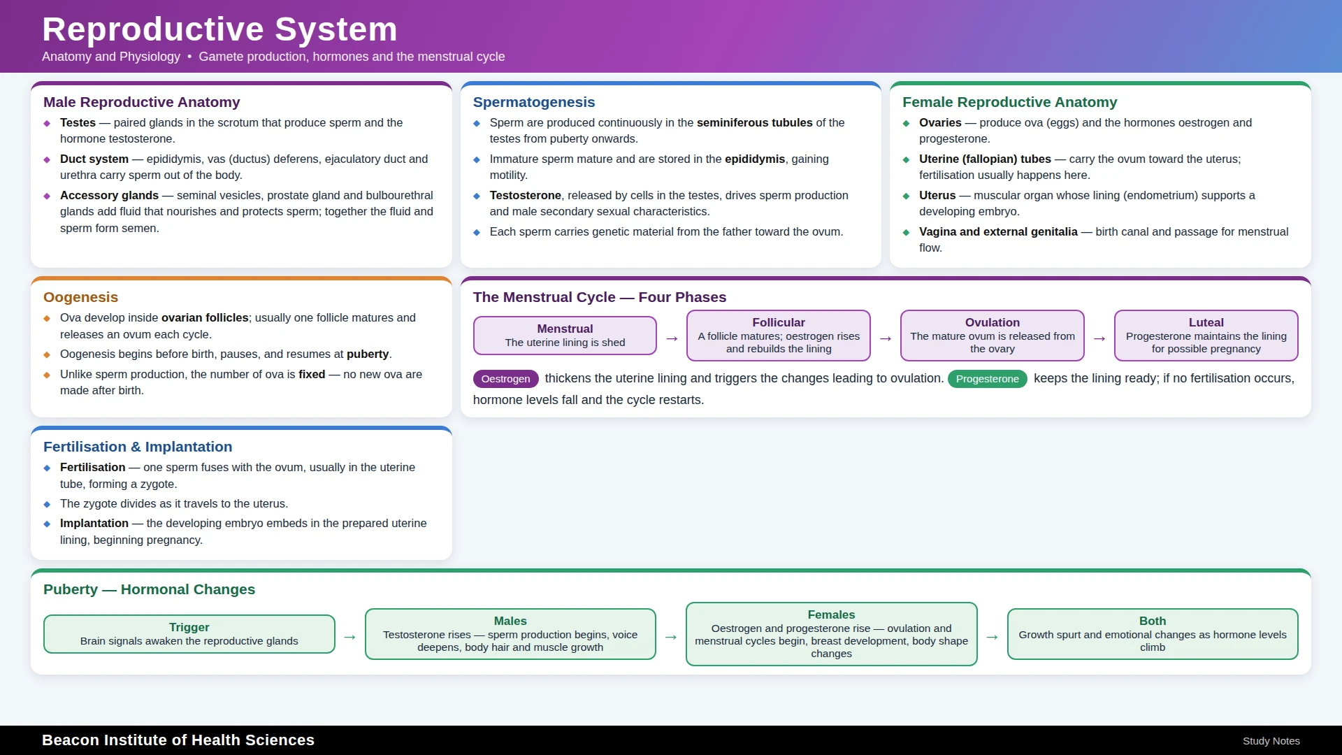
Gonads produce gametes and hormones: testes produce sperm and ovaries produce oocytes. Meiosis reduces chromosome number in developing gametes. Fertilization combines genetic material from gametes, ordinarily restoring the diploid number. Chromosome and developmental variations exist, so a simple diagram does not explain every individual. Distinguish production of a gamete from its maturation, transport and ability to participate in fertilization. These stages depend on different structures and coordinated physiological conditions.
3. Testes and scrotum
The testes contain seminiferous tubules involved in sperm production. Sertoli cells support developing germ cells, while Leydig cells produce testosterone in response to hormonal stimulation. The scrotum supports an environment important for testicular function, including temperature regulation. Testicular hormones also affect tissues beyond reproduction. Structure and function should be learned together. A student should not infer fertility from external appearance or perform examinations outside supervised training and consent requirements.
4. Sperm maturation and transport
Sperm pass from testicular channels toward the epididymis, where maturation and storage occur. The ductus deferens carries them onward; ejaculatory ducts connect with the urethral pathway. Trace the sequence rather than assuming sperm are fully mature at the moment of production. The urethra has both urinary and reproductive roles in typical testicular anatomy, but these functions occur under coordinated regulation. A pathway diagram should show ducts and direction clearly without confusing them with accessory glands.
5. Accessory glands and semen
Seminal vesicles, prostate and bulbourethral glands contribute secretions with different roles in the reproductive tract. Semen contains sperm and fluid components; semen volume does not directly establish sperm quality or fertility. The penis contains erectile tissues involved in vascular changes and sexual function. Erectile function, ejaculation and sperm production are related but distinct processes. Normal-physiology teaching should avoid presenting one function as a complete measure of reproductive health or making assumptions about personal behavior.
6. Ovaries and follicles
The ovaries contain follicles in which oocytes develop with supporting cells. Follicular development involves changing hormonal interactions, and ovulation releases an oocyte from a mature follicle. The remaining follicular tissue forms the corpus luteum, with important hormone production. Oocyte development differs from the continuing pattern of sperm production. Do not describe the ovary as simply a container that releases an identical new egg every month. Follicular recruitment, maturation and selection are coordinated processes.
7. Uterine tubes, uterus and vagina
Uterine tubes transport material between the ovarian region and uterus and are commonly the site of fertilization. The uterus includes endometrium, myometrium and outer covering. The cervix forms the lower region opening toward the vagina. The vagina and vulva are distinct terms: the vulva describes external genital structures. A labeled diagram should locate these structures and distinguish their functions. Clinical pain or bleeding cannot be diagnosed merely by matching the location to one anatomical organ.
8. Hypothalamic-pituitary-gonadal regulation
Gonadotropin-releasing hormone influences pituitary secretion of follicle-stimulating and luteinizing hormones. Their effects depend on the gonadal tissue and physiological context. Gonadal hormones and inhibin contribute to feedback. In testicular physiology, regulation supports testosterone production and spermatogenesis. In ovarian physiology, changing feedback patterns help coordinate follicular development and ovulation. Learn a complete axis with source, target and feedback rather than assuming FSH and LH have one identical action in every tissue.
9. Ovarian cycle
The ovarian cycle is commonly described as follicular phase, ovulation and luteal phase. Developing follicles contribute changing estrogen levels; the corpus luteum produces progesterone and other hormones. A sustained preovulatory hormonal pattern supports the LH surge associated with ovulation. Timing varies among people and cycles. A fixed day fourteen is a teaching simplification for an example cycle, not a reliable prediction for everyone or a safe basis for individual fertility decisions.
10. Uterine cycle
The uterine cycle involves menstrual, proliferative and secretory phases. The endometrium changes in response to hormonal patterns coordinated with the ovarian cycle. Estrogen supports proliferation, and progesterone helps secretory changes after ovulation. When pregnancy does not occur, changes in corpus-luteum function and hormone levels contribute to menstruation. Ovarian and uterine phases describe different structures even though they are connected. A useful comparison diagram places both timelines together and labels the source of each hormone.
11. Fertilization and early development
Fertilization ordinarily occurs in a uterine tube, followed by cell divisions during movement toward the uterus. Implantation is a later process in which the developing conceptus attaches within a suitable endometrial environment. Fertilization and implantation are not the same event. Early pregnancy involves new signaling and changing support for hormone production. This overview does not establish pregnancy location or viability. Clinical concerns require qualified assessment rather than interpreting symptoms through a classroom timeline.
12. Mammary function and life stages
Mammary structures include milk-producing glandular units and ducts. Prolactin supports milk production, while oxytocin supports milk ejection; these are separate physiological actions. Puberty, pregnancy, lactation and reproductive aging involve changing hormonal patterns. Menopause reflects a transition associated with ovarian function, but experiences vary and symptoms require individual consideration. Avoid assuming that every person has the same cycle, lactation experience or reproductive goal. Respectful care begins with asking rather than inferring.
13. Worked learning cases
First trace a sperm pathway from a seminiferous tubule to the urethral outlet, identifying maturation and added secretions. Next use a fictional cycle to relate a follicle, ovulation, corpus luteum and secretory endometrium without claiming fixed timing. Finally compare fertilization with implantation and milk production with milk ejection. These cases test distinctions between events that are often confused. Any real pain, abnormal bleeding or reproductive concern should be assessed through the appropriate clinical pathway.
14. Answered review and practical task
Create two labeled pathway diagrams and one ovarian-uterine comparison timeline. Answers: testes and ovaries are gonads; sperm mature in the epididymis; the endometrium is the uterine lining; ovulation releases an oocyte; fertilization and implantation are distinct; prolactin and oxytocin support different lactation functions. Why avoid fixed timing assumptions? Cycles vary. Why ask about preferences? Anatomy does not reveal personal goals. Recap: connect structure, transport, hormone regulation and physiological events while maintaining privacy and respectful language.