
Learning Objectives
This dedicated Anatomy and Physiology I unit aims to equip BSN students with a foundational understanding of the cardiovascular system. Upon completion, learners should be able to:
- Identify heart chambers, valves, and major vessels.
- Trace pulmonary, systemic, and coronary circulation.
- Explain cardiac conduction, the cardiac cycle, and cardiac output.
- Compare different types of blood vessels and components of blood.
- Relate these anatomical and physiological foundations to clinical observations of perfusion.
This module focuses on foundational anatomy and physiology and is not intended to be a repeat of nursing oxygenation or cardiovascular-disease management topics.
Heart Anatomy: Position, Coverings, and Wall Structure
The heart is centrally located in the thoracic mediastinum, with its apex directed inferolaterally towards the left side of the body.
Heart Coverings (Pericardium)
- Fibrous Pericardium: This is the tough, outer enclosure of the heart.
- Serous Pericardium: This layer has two parts: the parietal layer and the visceral layer. A thin, lubricating fluid occupies the space between these two layers.
- Visceral Pericardium (Epicardium): The visceral layer of the serous pericardium is also known as the epicardium.
Heart Wall Layers
The heart wall consists of three distinct layers:
- Epicardium: The outermost layer, synonymous with the visceral pericardium.
- Myocardium: The middle, muscular layer responsible for generating the contractile force necessary for pumping blood.
- Endocardium: The innermost layer lining the heart chambers.
The left ventricle's myocardial wall is notably thicker than that of the right ventricle. This structural difference is crucial because the systemic circulation, driven by the left ventricle, requires significantly higher pressure to perfuse the entire body compared to the lower pressure demands of pulmonary circulation.
Chambers, Valves, and the Pathway of Blood
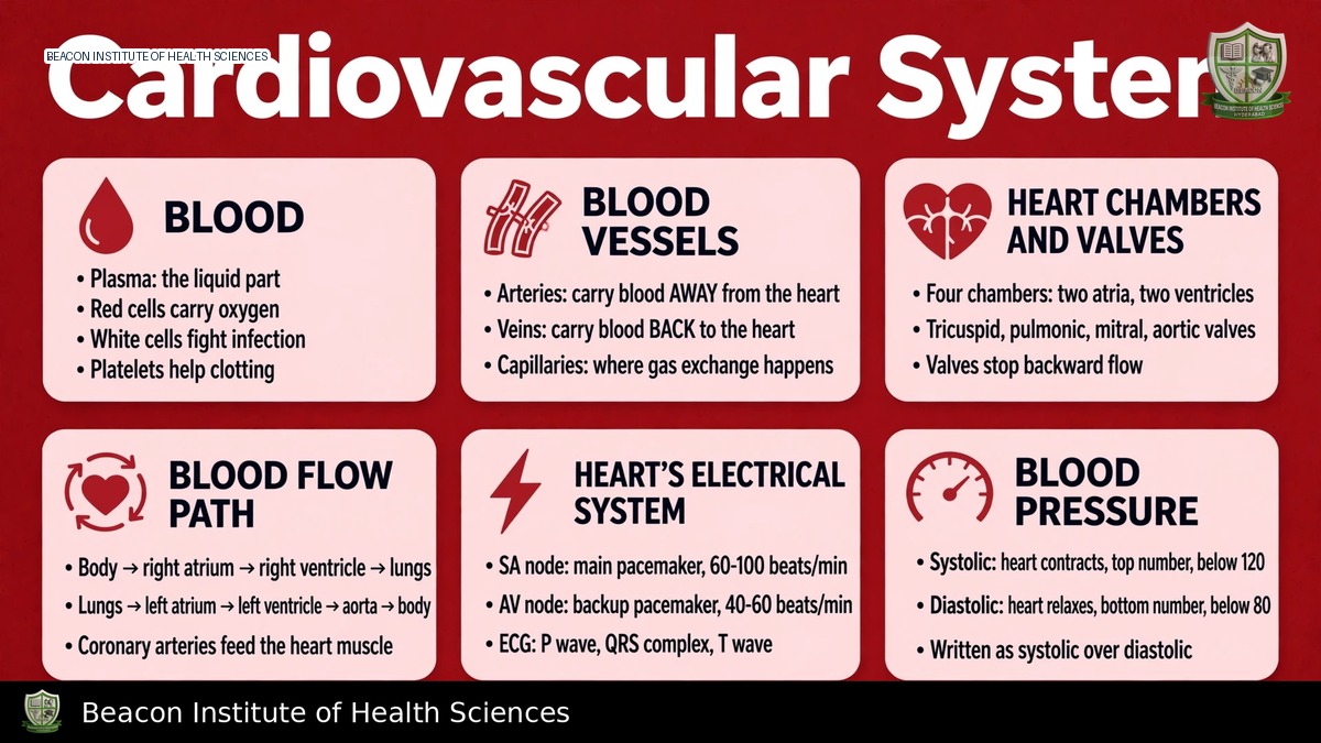
Blood circulates through a specific pathway within the heart's four chambers, regulated by a series of valves.
Blood Flow Through the Heart
- The right atrium receives systemic venous blood from the body via the superior and inferior venae cavae, as well as coronary venous blood from the coronary sinus.
- Blood then passes through the tricuspid valve into the right ventricle.
- From the right ventricle, blood is pumped through the pulmonary valve into the pulmonary trunk and arteries.
- In the lungs, gas exchange occurs, and the blood becomes oxygenated.
- Pulmonary veins deliver this oxygenated blood to the left atrium.
- The blood moves from the left atrium across the mitral (bicuspid) valve into the left ventricle.
- Finally, the left ventricle pumps the oxygenated blood through the aortic valve into the aorta, from where it is distributed to the rest of the body.
Key Definitions: Arteries, Veins, and Valve Function
- Arteries: Vessels that carry blood away from the heart. This definition is based on direction of flow, not oxygen content. For example, pulmonary arteries carry relatively deoxygenated blood, while systemic arteries carry oxygenated blood.
- Veins: Vessels that carry blood towards the heart. Pulmonary veins carry relatively oxygenated blood, while systemic veins carry deoxygenated blood.
- Atrioventricular (AV) Valves: The tricuspid and mitral valves are supported by chordae tendineae and papillary muscles, which prevent valve prolapse into the atria during ventricular contraction.
- Semilunar Valves: The pulmonary and aortic valves, unlike AV valves, do not possess chordae tendineae.
- All heart valves open and close primarily in response to pressure differences between chambers and vessels, rather than actively pumping blood.
The Three Circulatory Routes
The cardiovascular system involves three interconnected circulatory routes: pulmonary, systemic, and coronary circulation.
Pulmonary Circulation
This circuit connects the right side of the heart to the lungs and then back to the left side of the heart. It facilitates gas exchange, allowing blood to release carbon dioxide and pick up oxygen.
Systemic Circulation
Systemic circulation involves the left side of the heart, extends throughout the body's tissues, and returns to the right side of the heart. This route delivers oxygenated blood and nutrients to body tissues and carries deoxygenated blood and waste products away.
Coronary Circulation
Coronary arteries originate near the aortic root and are responsible for supplying oxygenated blood and nutrients directly to the myocardium (heart muscle). Coronary veins then return deoxygenated blood from the myocardium to the coronary sinus, which drains into the right atrium. It is important to note that blood within the heart chambers alone does not adequately nourish the entire heart wall. Myocardial oxygen delivery is a complex process influenced by coronary perfusion, pressure dynamics, and the metabolic demands of the heart, not solely by the blood oxygen level.
Electrical Conduction and ECG Foundations
The heart's rhythmic pumping action is coordinated by a specialized electrical conduction system.
Cardiac Conduction System
- The sinoatrial (SA) node, located in the right atrium, typically initiates atrial excitation, acting as the heart's natural pacemaker.
- Electrical signals spread through the atria and converge at the atrioventricular (AV) node, where they experience an important physiological delay.
- From the AV node, the signals pass through the atrioventricular bundle (Bundle of His), then divide into bundle branches, and finally reach the Purkinje network, which rapidly distributes the impulse throughout the ventricular myocardium to coordinate ventricular activation.
- Cardiac pacemaker activity is intrinsic, meaning the heart can generate its own electrical impulses.
Autonomic Influence on Heart Rate
The autonomic nervous system modulates heart rate and conduction:
- Sympathetic Activity: Generally increases heart rate and myocardial contractility.
- Parasympathetic Activity: Generally slows nodal activity, thus decreasing heart rate.
Electrocardiogram (ECG) Basics
The electrocardiogram (ECG) records the electrical activity of the heart at the body surface. Key waveforms correspond to specific electrical events:
- P wave: Represents atrial depolarization (electrical activation).
- QRS complex: Represents ventricular depolarization.
- T wave: Represents ventricular repolarization (electrical recovery).
It is critical to understand that electrical events recorded by the ECG precede the associated mechanical activity of the heart. An ECG waveform, while vital, does not directly prove effective cardiac output; a pulse assessment is necessary to ascertain actual circulation. Detailed ECG rhythm interpretation requires additional specialized training.
Cardiac Cycle and Heart Sounds
The cardiac cycle describes the sequence of mechanical and electrical events that occur during a single heartbeat, while heart sounds provide audible clues to valve function.
Phases of the Cardiac Cycle
- Diastole: This phase involves ventricular relaxation and subsequent filling with blood. Most ventricular filling occurs passively without atrial contraction under usual resting conditions, with atrial contraction contributing to the final portion of filling.
- Systole: This phase involves ventricular contraction and the ejection of blood into the pulmonary artery and aorta.
- Isovolumetric Contraction: During this brief period, all heart valves are closed, and ventricular pressure rises sharply without a change in ventricular volume.
- Ejection: This phase begins when ventricular pressure surpasses the outflow pressure in the great arteries, leading to blood expulsion from the ventricles.
- Following ejection, ventricular relaxation allows pressure to fall before the next filling phase resumes.
At an average heart rate of 75 beats per minute, one complete cardiac cycle lasts approximately 0.8 seconds. However, the duration of the cardiac cycle changes with variations in heart rate.
Heart Sounds (S1 and S2) and Murmurs
- First Heart Sound (S1): Primarily associated with the closure of the atrioventricular (tricuspid and mitral) valves, occurring near the beginning of ventricular systole.
- Second Heart Sound (S2): Primarily associated with the closure of the semilunar (aortic and pulmonary) valves, occurring near the beginning of diastole.
- Murmurs: These are sounds that reflect turbulent blood flow within the heart and require clinical interpretation. Not every extra or abnormal heart sound signifies a murmur.
Cardiac Output, Pressure, and Vascular System
Understanding cardiac output, blood pressure, and vessel characteristics is fundamental to cardiovascular physiology.
Cardiac Output Calculation and Determinants
Cardiac output (CO) is a measure of the volume of blood pumped by the heart per minute. It is calculated as:
- Cardiac Output (CO) = Heart Rate (HR) × Stroke Volume (SV)
As a classroom example, if the heart rate is 70 beats/min and the stroke volume is 70 mL/beat, the cardiac output would be approximately 4.9 L/min. It is important to note that this is an illustrative example, not a universal normal value.
Stroke volume, the amount of blood ejected by the ventricle with each beat, is influenced by three main factors:
- Preload: The degree of ventricular stretch before contraction.
- Contractility: The intrinsic strength of the ventricular muscle contraction.
- Afterload: The resistance the ventricles must overcome to eject blood.
An increased heart rate does not always lead to improved cardiac output, as excessively rapid rates can reduce the time available for ventricular filling, potentially decreasing stroke volume.
Blood Pressure Regulation
Blood pressure, the force exerted by blood against vessel walls, is influenced by several factors:
- Cardiac output
- Vascular resistance
- Circulating blood volume
- Properties of the blood vessels themselves
Structure and Function of Blood Vessels
- Arteries: Characterized by relatively thick, muscular walls, which enable them to withstand the high pressures generated by the heart.
- Arterioles: Smaller arteries that play a crucial role in regulating vascular resistance and, consequently, blood flow to specific tissues.
- Capillaries: These are microscopic vessels with extremely thin endothelial walls, facilitating efficient exchange of gases, nutrients, and waste products between blood and tissues.
- Veins: Often referred to as capacitance vessels due to their ability to hold a large volume of blood, veins have thinner walls than arteries. Many veins contain valves to prevent backflow of blood, and venous return to the heart is assisted by skeletal-muscle contractions and respiratory movements.
- Capillary Fluid Exchange: The movement of fluid between capillaries and interstitial spaces depends on a balance of hydrostatic and oncotic forces, as well as the barrier properties of the capillary walls. Lymphatic vessels are responsible for returning excess interstitial fluid and proteins to the circulation. It is important to note that not all filtered fluid returns directly to the capillary's venous end.
Blood Components and Nursing Implications
Blood is a vital fluid that performs numerous functions essential for life, and its properties have direct nursing implications.
Components and Functions of Blood
- Plasma: The liquid matrix of blood, carrying water, electrolytes, proteins, and various dissolved substances.
- Erythrocytes (Red Blood Cells): Primarily responsible for transporting oxygen throughout the body, facilitated by the protein hemoglobin.
- Leukocytes (White Blood Cells): Key components of the immune system, participating in defense against pathogens and foreign substances.
- Platelets: Essential for hemostasis (blood clotting) to prevent excessive bleeding.
In addition to these specific roles, blood as a whole transports gases, nutrients, waste products, and hormones. It also plays crucial roles in the body's regulation of temperature and pH, and in protection against infection and blood loss.
Clinical Relevance and Perfusion Assessment
BSN students should connect this anatomical and physiological knowledge to clinical assessments of perfusion. This includes observing pulse quality, trending blood pressure measurements, assessing skin temperature, evaluating consciousness, and monitoring urine output. Adequate tissue oxygen delivery relies on both sufficient blood flow and oxygen content; therefore, a satisfactory oxygen saturation level alone does not guarantee adequate perfusion to tissues. Nurses should escalate concerning chest symptoms or any signs of compromised circulation according to local protocols. A practical exercise for revision involves tracing one complete blood circuit, matching valves with chambers, and clearly differentiating between electrical conduction, ECG activity, and mechanical pumping.