
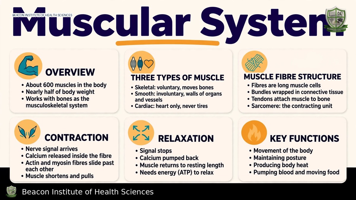
Learning Objectives and Functions of the Muscular System
This lesson aims to help BSN students and nursing educators compare skeletal, cardiac, and smooth muscle, explain skeletal muscle organization and contraction, identify major muscle groups, and relate muscle function to movement, posture, heat production, and nursing assessment.
Core Properties of Muscle Tissue
Muscle tissue possesses several key properties:
- Excitability: The ability to respond to stimuli.
- Contractility: The ability to shorten forcefully.
- Extensibility: The ability to stretch without being damaged.
- Elasticity: The ability to recoil to original length after stretching.
Skeletal muscles also play crucial roles in stabilizing joints and supporting selected body openings and soft tissues.
Three Primary Muscle Types
Skeletal Muscle
- Features long, multinucleated, striated fibers.
- Primarily controlled by somatic motor neurons.
- Responsible for voluntary movement, though reflexes can also activate it.
Cardiac Muscle
- Composed of branched, striated cells, typically with one central nucleus.
- Cells are connected by intercalated discs, facilitating coordinated involuntary activity to pump blood.
Smooth Muscle
- Consists of non-striated, spindle-shaped cells with a single nucleus.
- Found in the walls of hollow organs and vessels.
- Responds to autonomic, hormonal, and local signals.
- It is important to note that striation does not automatically imply voluntary control.
From Whole Muscle to Sarcomere: Organization and Structure
A whole skeletal muscle contains not only muscle fibers but also supporting connective tissue, blood vessels, and nerves.
Connective Tissue Layers
- Epimysium: Surrounds the entire muscle.
- Perimysium: Surrounds bundles of muscle fibers called fascicles.
- Endomysium: Surrounds individual muscle fibers.
Tendons or broad aponeuroses connect many muscles to their attachments.
The Muscle Fiber and Its Components
- Sarcolemma: The muscle-fiber membrane.
- Sarcoplasm: The cytoplasm within the muscle fiber.
- Myofibrils: Contain repeating contractile units called sarcomeres, which are located between Z discs.
- Thick filaments: Composed of myosin.
- Thin filaments: Composed of actin, along with troponin and tropomyosin.
Sarcomere Structure: Bands and Zones
- A band: Spans the thick filaments, including regions where thick and thin filaments overlap.
- I band: Contains only thin filaments.
- H zone: Contains only thick filaments without thin overlap.
- T tubules: Carry membrane excitation inward from the sarcolemma.
- Sarcoplasmic Reticulum: Stores and releases calcium ions.
- A skeletal-muscle triad includes a T tubule flanked by terminal cisternae on both sides.
Neuromuscular Transmission
When a motor-neuron impulse reaches the nerve terminal, it promotes the release of acetylcholine. Acetylcholine then binds to receptors at the motor end plate, causing local depolarization. If this depolarization reaches threshold, it initiates a muscle action potential. This excitation spreads over the sarcolemma and T tubules, leading to the release of calcium from the sarcoplasmic reticulum. Acetylcholinesterase limits the action of acetylcholine. The neuromuscular junction serves to transfer a signal; it is not a direct physical continuity between the neuronal and muscle cytoplasm.
Sliding-Filament Contraction and Relaxation
Mechanism of Contraction
Muscle contraction begins when calcium binds to troponin, causing tropomyosin to shift and expose actin binding sites. Energized myosin heads then attach to actin, perform a power stroke, and detach when ATP binds. The hydrolysis of ATP prepares the myosin head for another cycle. As this cycle repeats, thin filaments slide past thick filaments, bringing the Z discs closer. The filaments themselves do not shorten. During this process, the I band and H zone narrow, while the A band remains broadly unchanged.
Mechanism of Relaxation
When neural stimulation ceases, calcium is actively pumped back into the sarcoplasmic reticulum using energy. This allows the binding sites on actin to be covered again by tropomyosin, and muscle tension declines. ATP is essential for both cross-bridge detachment and calcium handling. A lack of ATP does not immediately relax the muscle fiber. These mechanisms explain normal muscle activity but do not serve as a treatment model for electrolyte or neurological diseases.
Force Generation, Energy, and Contraction Types
Motor Units and Force Regulation
A motor unit comprises a motor neuron and all the muscle fibers it innervates. The force generated by a muscle can be increased by recruiting additional motor units and by increasing the frequency of stimulation.
- A twitch is a brief, single muscle response.
- Temporal summation and tetanic contraction result from appropriate repeated stimulation. In this context, tetanus refers to a sustained physiological contraction, distinct from infectious tetanus.
Types of Muscle Contraction
- Isometric contraction: Develops tension without significant change in muscle length, such as holding a posture.
- Concentric contraction: Shortens a muscle while producing force.
- Eccentric contraction: Lengthens a muscle under tension, for instance, during controlled lowering.
Muscle tone refers to the low-level, ongoing activation of muscles that helps support posture.
Energy Sources for Muscle Activity
Muscle energy is derived from available ATP, phosphocreatine, anaerobic glycolysis, and aerobic metabolism. The proportions in which these sources are used vary depending on the duration and demand of the activity. Muscle fatigue has multiple peripheral and central causes and is not solely explained by lactate accumulation.
Muscle Actions and Major Muscle Groups
Functional Roles of Muscles
- An agonist is the principal mover of a movement.
- An antagonist opposes a movement.
- Synergists or fixators assist and stabilize.
- Origin and insertion typically describe relatively fixed and more mobile attachments, respectively, though their functional roles are task-dependent.
Key Muscle Groups and Their Actions
- Biceps brachii: Supports elbow flexion and forearm supination.
- Triceps brachii: Extends the elbow.
- Deltoid: Supports shoulder abduction.
- Pectoralis major: Acts at the shoulder.
- Trapezius and other muscles: Control scapula position.
- Diaphragm: The main inspiratory muscle.
- Intercostals: Assist thoracic movement.
- Abdominal muscles: Support the trunk and abdominal pressure.
- Spinal extensors: Help maintain posture.
- Gluteus maximus: Extends the hip.
- Gluteus medius: Contributes to pelvic stability.
- Quadriceps: Extend the knee.
- Hamstrings: Generally flex the knee and help extend the hip.
- Gastrocnemius and soleus: Plantar-flex the ankle.
- Tibialis anterior: Dorsiflexes the ankle.
For effective study, it is recommended to learn each muscle action in conjunction with its joint, rather than memorizing names alone.
Training, Disuse, and Nursing Relevance
Factors Affecting Muscle Function
Regular loading can increase skeletal muscle-fiber size and performance; however, adult strength gains typically do not result primarily from the production of many new fibers. Conversely, disuse leads to muscle atrophy and weakness. Recovery after injury depends on the type and extent of damage, and muscle function is also affected by aging, nutrition, neural input, and illness.
Nursing Assessment Considerations
Nurses should observe strength, symmetry, tone, movement, and tolerance within their clinical scope. It is crucial to distinguish joint restriction from actual muscle weakness and pain-limited effort. Safe patient transfers, mobilization, and breathing assessments depend on coordinated muscle activity. Prescribed precautions must always govern actual exercises.
Key Revision Points for Nurses
To reinforce understanding, students should revise by tracing an impulse from a motor neuron to a cross-bridge, explaining the various roles of ATP, and comparing isometric, concentric, and eccentric muscle actions using a simple movement.