
Learning Objectives
This lesson aims to equip BSN students with a comprehensive understanding of human joints. Upon completion, you should be able to:
- Classify joints based on their structure and function.
- Describe the key features of a synovial joint.
- Identify common joint types and their characteristic movements.
- Explain how joint mobility and stability are critical in supporting nursing care.
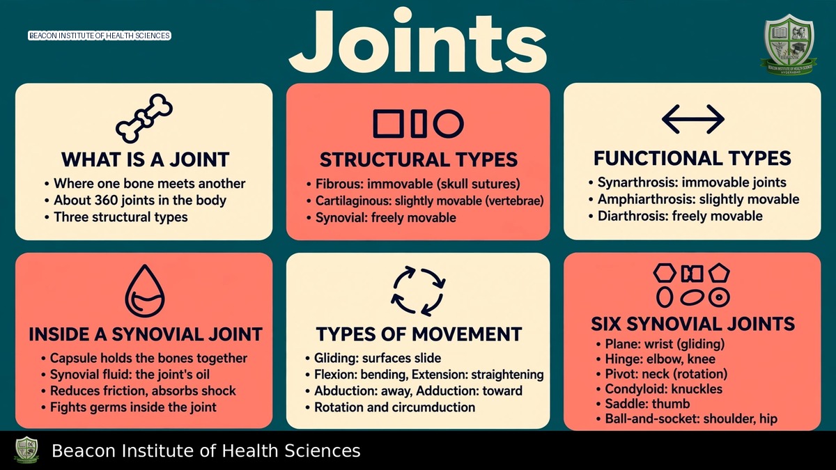
An articulation is fundamentally a connection between skeletal structures. These connections vary greatly, with some allowing minimal movement and others supporting extensive movement. It's crucial to understand that structural and functional classifications address different aspects and should not be conflated.
Structural Classification of Joints
Joints are categorized structurally based on the material binding the bones and the presence or absence of a joint cavity.
Fibrous Joints
Fibrous joints are characterized by bones united by fibrous connective tissue, lacking a joint cavity. Examples include:
- Sutures: Typical examples are the skull sutures.
- Syndesmoses: These involve ligaments or interosseous membranes, such as the distal tibiofibular connection, allowing limited movement.
- Gomphoses: These joints anchor teeth within their sockets via periodontal ligaments.
Cartilaginous Joints
Cartilaginous joints also lack a synovial cavity. Their articulating bones are united by cartilage. Types include:
- Synchondroses: Utilize hyaline cartilage, as seen in growth plates and the first sternocostal joint.
- Symphyses: Involve fibrocartilage, exemplified by the pubic symphysis and the articulations between vertebral bodies. It's important to note that the first sternocostal joint differs from many other rib-sternum articulations, and not every rib connection should be classified identically.
Synovial Joints
Synovial joints feature an enclosed fluid-containing cavity, allowing for greater movement. Common examples include the shoulder, elbow, hip, and knee. The stability of these joints is maintained by the arrangement of ligaments, the joint capsule, their shape, and the controlling muscle action.
Functional Classification of Joints
Functional classification categorizes joints by the amount of movement they permit:
- Synarthrosis: These joints are essentially immovable. Most sutures are synarthroses.
- Amphiarthrosis: These joints allow limited movement. Symphyses commonly fall into this category.
- Diarthrosis: These joints are freely movable. All synovial joints are diarthroses; however, this does not imply that every synovial joint moves in all directions.
A functional description should always be paired with its specific structural classification rather than memorized as a perfect one-to-one mapping for all fibrous and cartilaginous joints.
Synovial Joint Structure
The intricate structure of synovial joints facilitates movement and distributes load:
- Articular Hyaline Cartilage: Covers the opposing bone surfaces, supporting low-friction load transfer. This cartilage is avascular.
- Fibrous Capsule: Encloses the joint.
- Internal Synovial Membrane: Lines appropriate non-cartilaginous surfaces within the capsule.
- Synovial Fluid: Lubricates the joint and supplies nutrients to the avascular cartilage. The membrane itself does not cover the articular cartilage.
- Ligaments: Connect bone to bone, restraining excessive motion.
- Tendons: Transmit muscle force to bone. Ligaments and tendons, despite similar connective tissue composition, have distinct functions.
- Accessory Structures: Some joints feature fibrocartilaginous discs or menisci, which enhance congruence and distribute load. Bursae reduce friction between moving tissues, and tendon sheaths support tendon movement. These structures are not uniformly present in all joints, and a bursa is distinct from the main joint cavity.
Six Common Synovial Joint Types
Synovial joints are further classified by the shape of their articulating surfaces, which dictates their range of motion:
- Plane Joints: Permit gliding movements, such as many intercarpal joints.
- Hinge Joints: Primarily allow flexion and extension, exemplified by the elbow. The knee is considered a modified hinge joint.
- Pivot Joints: Permit rotation around an axis, like the proximal radioulnar joint.
- Condyloid or Ellipsoid Joints: Allow movement in two main planes, such as the radiocarpal wrist joint.
- Saddle Joints: Exemplified by the thumb's first carpometacarpal articulation, enabling movements vital for opposition.
- Ball-and-Socket Joints: Permit multiaxial movement, such as the shoulder and hip. The shoulder joint favors mobility, while the deeper hip socket and strong supports prioritize weight-bearing stability. It is important not to assume stability or mobility solely based on the joint type.
Movement Terminology
Precise terminology is essential for describing joint movements:
- Flexion: Generally decreases the angle between segments.
- Extension: Increases the angle between segments.
- Hyperextension: Extends a joint beyond its anatomical position where possible.
- Abduction: Moves a limb away from the body's reference midline.
- Adduction: Moves a limb toward the body's reference midline.
- Circumduction: Combines movements so the distal segment traces a cone; it is not the same as rotation.
- Rotation: Turns a structure about an axis, with internal/medial and external/lateral directions.
- Pronation: Positions the forearm so the radius rotates relative to the ulna.
- Supination: Returns the palm forward to the anatomical position.
- Dorsiflexion: Raises the front of the foot toward the leg.
- Plantar Flexion: Points the foot downward.
- Inversion: Turns the sole of the foot inward.
- Eversion: Turns the sole of the foot outward.
- Elevation: Moves a structure upward.
- Depression: Moves a structure downward.
- Protraction: Moves a structure forward.
- Retraction: Moves a structure backward.
- Thumb Opposition: Brings the thumb toward finger pads.
- Reposition: Returns the thumb to its anatomical position.
Always use the correct body region when documenting movement.
Stability, Cartilage, and Ageing
Joint stability is maintained by both passive and active mechanisms. Congruent joint surfaces, the capsule, and ligaments provide passive stability, while muscles contribute to active control. Articular cartilage, lacking a direct blood supply, has limited healing capacity. Joint nutrition and function are supported by movement and appropriate loading. Immobilization can reduce mobility and lead to contracture, necessitating careful consideration of injuries, surgery, and prescribed restrictions for movement. Age-related changes and degenerative joint disease differ from normal joint anatomy, and inflammation can cause pain, swelling, and functional limitation without identifying a single cause.
Nursing Application and Revision
For BSN students and practicing nurses, understanding joints is critical for patient care:
- Assessment: Assess active movement (performed by the person) and passive movement (guided by another) only when appropriate and within scope.
- Documentation: Accurately record the affected joint, side, movement, limitations, and symptoms.
- Safety: Never force a painful joint or assume a standard exercise is safe after injury. Fall prevention, safe transfers, and proper positioning rely on both joint mobility and muscle control.
- Prompt Intervention: Suspected dislocation or an acute neurovascular problem requires immediate escalation, not classroom maneuvers.
To reinforce your knowledge, practice these revision exercises:
- Classify a skull suture, pubic symphysis, and shoulder joint using both structural and functional systems.
- Draw and label a typical synovial joint.
- Demonstrate forearm pronation and supination, distinguishing it from shoulder rotation.
- Explain why ligaments and tendons have differing functions despite similar connective tissue composition.
- Discuss how extensive joint mobility can sometimes reduce intrinsic stability.