
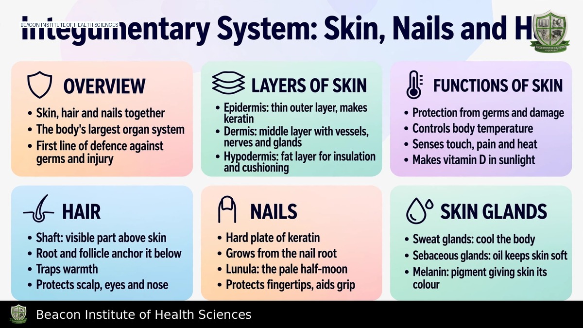
Reference-based BSN study notes for Anatomy and Physiology I.
Learning objectives and scope
Describe skin layers and cell types; explain hair, nails and glands; and connect barrier, sensory and thermoregulatory functions to nursing observations. This Anatomy and Physiology I unit examines normal structure and function. It differs from a skin-care, hair-loss or wound-treatment article even though some clinical examples overlap.
System functions
Skin and its appendages protect against physical injury, excessive water loss and many environmental exposures. Surface lipids, epithelial integrity and local immune activity contribute to protection. Sensory receptors detect touch, pressure, temperature and pain. Cutaneous blood flow and sweating assist thermoregulation. Ultraviolet exposure initiates vitamin D synthesis in skin, followed by metabolic steps elsewhere; this is not a recommendation for unsafe sun exposure. Skin also provides a visible surface for observing perfusion and tissue changes.
Epidermis
The epidermis is keratinised stratified squamous epithelium and has no blood vessels of its own. It receives nutrients by diffusion from underlying tissue. Keratinocytes form most cells and contribute to the barrier. Melanocytes produce melanin, which is transferred to keratinocytes and assists ultraviolet protection. Epidermal dendritic/Langerhans cells participate in immune surveillance, while tactile/Merkel cells contribute to touch perception. Skin colour reflects melanin, haemoglobin and other influences, so colour alone is not a reliable measure of oxygenation or perfusion.
From deep to superficial, epidermal strata are basale, spinosum, granulosum and corneum, with lucidum between granulosum and corneum in thick skin. Basal cells divide and replace cells moving toward the surface. The outer corneum contains flattened keratinised cells contributing to protection. Thick skin on palms and soles has the additional lucidum and lacks hair follicles and sebaceous glands. Thin skin covers most other areas and can contain hair and sebaceous glands. Thick skin refers primarily to epidermal thickness, not simply a thick subcutaneous fat layer.
Dermis and hypodermis
The dermis is connective tissue containing collagen, elastic fibres, vessels, nerves and appendage structures. Its superficial papillary layer includes loose connective tissue and dermal papillae; the deeper reticular layer is dense irregular connective tissue. Dermal organisation supports strength, flexibility and sensory function. Epidermal ridges and dermal interactions contribute to surface patterns.
The hypodermis or subcutaneous layer lies beneath the skin and contains variable amounts of adipose and loose connective tissue. It cushions, insulates and anchors skin while providing energy storage. Strict anatomical descriptions define skin as epidermis plus dermis; the hypodermis is closely associated but not one of those two layers. Layer terminology matters when discussing injuries and routes of injection, but selecting injection sites and depths requires clinical protocols rather than this overview.
Hair
A hair shaft projects above the surface; the root lies within a follicle. Growth originates from proliferating cells in the bulb's matrix, supported by the vascular dermal papilla. Keratinisation produces the shaft, with cuticle, cortex and sometimes medulla. Hair supports protection and sensation in different locations. Arrector pili are smooth muscles attached to follicles and produce goosebumps; sebaceous glands commonly open into follicles.
Hair cycles through growth, transition and resting phases, often termed anagen, catagen and telogen. Follicles are not all synchronised, so normal shedding occurs alongside growth. Nutrition, hormones, illness and medications can influence the cycle, but a change in hair alone does not establish a diagnosis.
Nails
The nail plate is a hard keratinised structure over the nail bed. The matrix produces most new nail plate, and the root lies proximally beneath the skin fold. The lunula is the visible pale crescent of the proximal matrix in some nails. The eponychium/cuticle and surrounding folds help protect the proximal region; the hyponychium is beneath the free edge. Nails protect distal digits and support precise manipulation. The plate itself lacks nerves and vessels, whereas surrounding living tissues have them.
Nail colour, shape and growth can change for local or systemic reasons. These findings require context and assessment; they should not be used alone to diagnose nutritional deficiency, hypoxia or infection.
Cutaneous glands
Eccrine sweat glands open onto the surface and support heat loss through evaporation. Apocrine glands occur in selected regions and commonly open into follicles; their activity becomes more prominent after puberty. Odour often results when microbes metabolise secretions, rather than fresh secretion inherently having the final odour. Sebaceous glands produce sebum, supporting lubrication of skin and hair. Ceruminous and mammary glands are specialised glandular structures discussed in relevant regional units. Sweating contributes limited waste elimination but is not the body's main detoxification pathway.
Repair, ageing and nursing application
Superficial injury can heal through epithelial regeneration, whereas deeper damage may require connective-tissue repair and scar formation. Healing involves haemostasis, inflammation, proliferation and remodeling; phases overlap. Ageing can reduce elasticity, skin thickness and protective reserve, while perfusion, moisture, nutrition and pressure influence integrity. Avoid assuming all redness or pigmentation has the same meaning across skin tones.
Inspect skin, hair and nails systematically and document location, integrity, moisture, temperature and relevant changes. Pressure and shear can injure tissues even before a surface wound is obvious. Assess findings alongside the patient's wider condition and follow local prevention and escalation protocols. Revise by tracing layers from surface to subcutaneous tissue, distinguishing hair matrix from hair shaft, and explaining why a deep injury heals differently from a superficial abrasion.