
1. Learning objectives and scope
The nervous system detects information, integrates it and coordinates responses. In Anatomy and Physiology II, BSN students should connect structures with functions rather than memorize names alone. This lesson covers neurons, major divisions, brain and spinal cord organization, signaling, reflexes and autonomic control. Clinical examples illustrate concepts and do not replace supervised assessment, diagnosis or local emergency procedures. A symptom can have several causes and cannot establish a neurological diagnosis by itself.
2. Central and peripheral organization
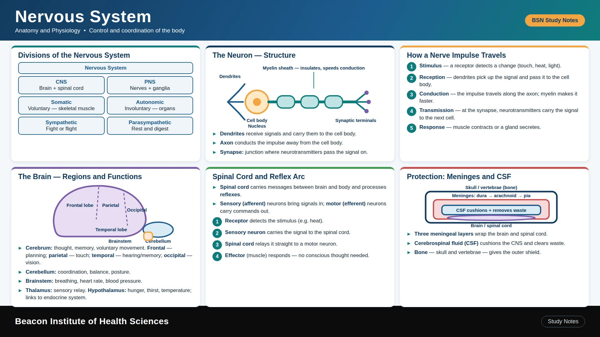
The central nervous system, or CNS, consists of the brain and spinal cord. The peripheral nervous system, or PNS, includes nerves and ganglia that connect central structures with the body. Sensory, or afferent, information travels toward the CNS; motor, or efferent, commands travel toward effectors. These directions are defined relative to the CNS. A mixed nerve contains both sensory and motor fibers, so one nerve can support more than one kind of function.
3. Neuron structure
A neuron has a cell body, dendrites and an axon. Dendrites usually receive inputs; the axon carries signals toward its terminals. Neurons vary in shape and function. Sensory neurons carry information from receptors, motor neurons supply effectors and interneurons participate in processing. The cell body maintains the cell, while communication depends on membrane properties and connections. A useful study diagram should label the cell body, nucleus, dendrites, axon and terminal without implying that every neuron has identical anatomy.
4. Glia and myelin
Glial cells support nervous tissue in several ways. Oligodendrocytes form CNS myelin, and Schwann cells form PNS myelin. Myelin helps efficient conduction along many axons; gaps are called nodes of Ranvier. Astrocytes support the local environment, microglia participate in immune responses and ependymal cells line ventricular spaces. Myelin is not present on every axon. Damage to neural tissue can affect communication, but the effect depends on the location and extent of injury.
5. Membrane potential and action potentials
Ion concentration differences and selective membrane permeability contribute to a resting membrane potential. When sufficient stimulation reaches threshold, an action potential involves rapid changes in ion movement across the membrane. It is an all-or-none event; stronger stimuli do not simply produce larger action potentials. Signal frequency and the number of active neurons contribute to information coding. Refractory periods help organize signaling. Do not confuse electrical events along an axon with the chemical transmission occurring at many synapses.
6. Synapses and neurotransmission
At a chemical synapse, an arriving action potential promotes neurotransmitter release. The transmitter interacts with receptors on the next cell and changes its activity. Some effects are excitatory and others inhibitory, depending on the transmitter, receptor and context. Signals may end through reuptake, breakdown or diffusion. Neural networks integrate many inputs rather than operating as a single straight wire. This explains why a change at one connection does not automatically predict the behavior of the whole system.
7. Cerebrum and functional areas
The cerebrum contains two hemispheres connected by pathways including the corpus callosum. The cortex participates in perception, voluntary movement, language, reasoning and memory through distributed networks. Frontal, parietal, temporal and occipital lobes provide useful anatomical landmarks. Motor and sensory functions often show contralateral organization, but this is not a universal rule for every function. Avoid simplistic statements that one hemisphere alone is logical or creative. Complex activities depend on interacting regions.
8. Diencephalon, brainstem and cerebellum
The thalamus is an important relay and processing region for many sensory pathways. The hypothalamus links neural control with homeostasis and endocrine regulation. The brainstem includes midbrain, pons and medulla and contains pathways and nuclei involved in essential functions. The cerebellum supports coordination, balance and motor learning. These regions work together. An educational diagram should identify their locations and principal roles without suggesting that one structure independently controls all aspects of consciousness or movement.
9. Protection and cerebrospinal fluid
The skull, vertebral column and meninges provide protection. The meningeal layers are dura mater, arachnoid mater and pia mater. Cerebrospinal fluid supports and cushions CNS structures and circulates through ventricular and subarachnoid spaces. The blood-brain barrier restricts movement of many substances but is not an absolute shield against every drug, organism or toxin. Understanding protection helps explain why pressure or inflammation can have important effects, while actual assessment requires trained clinical evaluation.
10. Spinal cord, roots and pathways
The spinal cord conducts information and participates in reflexes. Gray matter contains many cell bodies and local connections; white matter contains major axonal pathways. Dorsal roots carry sensory fibers and contain associated sensory ganglia; ventral roots carry motor fibers. Ascending pathways carry information toward higher centers, while descending pathways convey motor control. A spinal nerve forms from the roots and is mixed. Trace a pathway carefully rather than using dorsal and sensory as interchangeable labels for every structure.
11. Reflex organization
A reflex is a relatively rapid response to a stimulus through a neural circuit. A basic arc includes receptor, sensory neuron, integration center, motor neuron and effector. Some reflexes involve fewer connections than others. Withdrawal from a painful stimulus can begin through spinal processing, while information also reaches the brain for perception. Reflexes are not all confined to the spinal cord. A classroom demonstration must be safe, consented and supervised; students should not use painful stimulation as a home experiment.
12. Somatic and autonomic control
Somatic motor pathways supply skeletal muscle. Autonomic pathways influence smooth muscle, cardiac muscle and glands, commonly using a preganglionic and postganglionic neuron. Sympathetic and parasympathetic divisions support different patterns of regulation; they are not always simple opposites. The enteric system organizes substantial gastrointestinal activity while interacting with autonomic inputs. A faster heartbeat during a challenge illustrates coordinated control, but a heart-rate change alone does not identify a neurological disorder.
13. Worked learning case and nursing relevance
In a fictional classroom case, a person touches an unexpectedly hot object and pulls away before describing the pain. Trace receptor activation, sensory input, spinal integration, motor output and muscle contraction, then explain conscious perception through higher pathways. A second exercise maps the route of a voluntary hand movement from central planning to muscle activation. These cases separate reflex response from awareness. In practice, sudden weakness, altered consciousness or other acute changes require prompt assessment through local procedures, not diagnosis from a study diagram.
14. Practical task and answered review
Draw CNS and PNS divisions, label a neuron and compare sensory with motor direction. Add a five-part reflex arc and summarize somatic versus autonomic effectors. Answers: the CNS is brain and spinal cord; afferent means toward the CNS; oligodendrocytes form CNS myelin; a mixed nerve carries sensory and motor fibers; the cerebellum supports coordination. A correct diagram should show connections and direction, not only labels. Recap by connecting a structure, a function and a safe learning example.