
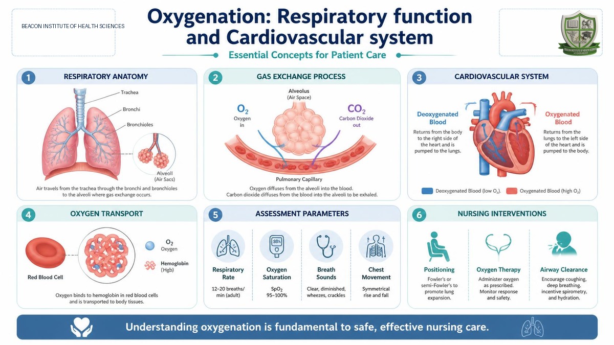
Factors Interfering with Effective Tissue Oxygenation
Oxygenation is a dynamic physiological process that can be altered by various multi-dimensional factors, categorized into environmental, lifestyle, health status, and gender factors.
Environmental Factors
- High Altitude: As atmospheric altitude increases, the partial pressure and density of oxygen in the air decrease. Less oxygen is inhaled with each breath, leading the body to compensate by increasing both the respiratory rate and heart rate to satisfy metabolic oxygen demands.
- Heat (High Temperature): Elevated temperatures cause superficial vasodilation of peripheral blood vessels, which decreases peripheral vascular resistance. This leads to faster blood flow, increased cardiac output, more vigorous heartbeats, and a rise in myocardial oxygen demand.
- Cold (Low Temperature): Exposure to cold triggers peripheral vasoconstriction, reducing cardiac output and lowering both heart and respiratory rates.
- Air Pollution: Gaseous or particulate air pollutants cause severe bronchial irritation and bronchospasm, inducing significant breathing difficulties (dyspnea) in individuals with a history of underlying lung diseases or altered respiratory baselines.
Lifestyle Factors
- Physical Activity: Systematic physical exercise increases metabolic rates, requiring the respiratory system to increase the rate and depth of breathing, which correspondingly elevates the heart rate.
- Sedentary Lifestyle: Lack of physical exercise decreases metabolic oxygen demands, leading to a shallow breathing pattern, lower respiration rates, and slower peripheral blood circulation.
Health Status and Medications
- Underlying Diseases: Pre-existing cardiovascular or respiratory illnesses (such as atherosclerosis, chronic bronchitis, or emphysema) directly restrict gas exchange, compromise cardiac pumping, and prevent adequate tissue oxygenation.
- Anemia: A critical hematological condition characterized by a severe lack of red blood cells (RBCs) or hemoglobin, preventing sufficient oxygen binding and transport despite adequate lung ventilation.
- Narcotics and CNS Depressants: Medications used for severe pain relief, such as narcotics (including Morphine and Pethidine), directly depress the central nervous system. Specifically, they depress the respiratory center in the medulla oblongata, significantly lowering the respiratory rate and depth, which can lead to life-threatening respiratory arrest.
- Stress and Coping: Psychological stress triggers the sympathetic nervous system ('fight-or-flight' response), elevating heart and respiratory rates. Effective coping mechanisms activate the parasympathetic system, restoring cardiorespiratory rates to normal resting baselines.
Gender Factors
- Estrogen Protection: Prior to menopause, females produce high levels of estrogen, which provides a natural cardiovascular shield, actively protecting blood vessels against atherosclerosis and coronary artery diseases. Consequently, pre-menopausal women have a significantly lower incidence of cardiovascular illnesses compared to age-matched males.
Manifestations of Altered Respiratory and Cardiovascular Function
When cardiorespiratory function is compromised, several distinct clinical signs and patterns manifest across the systems.
Clinical Definitions in Oxygenation Alterations
- Hypoxia: A critical physiological state characterized by insufficient oxygen delivery to the body tissues at the cellular level. It can arise from ventilatory failure, alveolar gas diffusion barriers, or impaired circulatory transport.
- Hypoxemia: An abnormally low concentration of oxygen specifically in arterial blood. While hypoxemia refers strictly to blood gas values, hypoxia describes tissue-level oxygen deprivation.
- Cyanosis: A bluish discoloration of the skin, nail beds, and mucous membranes. It is associated with severe hypoxia and is caused by an accumulation of reduced (deoxygenated) hemoglobin in the superficial microvascular bed.
Clinical Signs and Symptoms of Tissue Hypoxia
- Neurological: Rapidly developing restlessness, severe anxiety, and lightheadedness.
- Respiratory: Rapid and shallow breathing, nasal flaring (active flaring of nostrils during breathing), and substernal/intercostal muscle retractions (visible pulling inward of the chest wall and rib spaces during inhalation).
- Cardiovascular: Tachycardia and eventual cyanosis as deoxygenated blood pools in superficial capillary networks.
Classification of Altered Breathing Patterns
Breathing patterns can be classified clinically based on Rate, Volume, Rhythm, and Effort:
| Category | Pattern | Clinical Description & Characteristics |
|---|---|---|
| Based on Rate | Tachypnea | Abnormally rapid respiration rates, typically presenting with shallow breathing. |
| Based on Rate | Bradypnea | Abnormally slow respiration rates, falling below the normal clinical baseline. |
| Based on Rate | Apnea | The temporary or complete cessation of breathing (absence of respiration). |
| Based on Volume | Hyperventilation | Deep, rapid breathing with prolonged inhalation, increasing air volume in the lungs. |
| Based on Volume | Hypoventilation | Shallow, abnormally slow respiration, reducing the volume of air reaching the alveoli. |
| Based on Rhythm | Cheyne-Stokes | Progressive increase in rate and depth, followed by gradual slowing, culminating in a temporary period of apnea (repeats every 30 seconds to 2 minutes). |
| Based on Effort | Dyspnea | Difficult, labored, or painful breathing, where the patient experiences intense 'air hunger'. |
| Based on Effort | Orthopnea | Dyspnea that occurs when lying flat; the client can only breathe comfortably in an upright sitting or standing position. |
Altered Cardiovascular Manifestations
- Decreased Cardiac Output: Occurs when myocardial blood flow is blocked due to atherosclerosis (fatty plaque accumulation) or thrombi (blood clots) in the coronary arteries. Complete obstruction leads to myocardial ischemia and cell death, known as Myocardial Infarction (MI) or a heart attack. If damage involves a large portion of the myocardium (especially the left ventricle), the heart fails to contract adequately, leading to Heart Failure.
- Impaired Tissue Perfusion: Arterial plaques (atherosclerosis) narrow vessels, reducing arterial flow to peripheral organs. Under-perfused tissues experience Ischemia (inadequate blood supply). Temporary ischemia in the heart causes Angina Pectoris (temporary chest pain); in the brain, it causes a Transient Ischemic Attack (TIA).
- Blood Alterations: Abnormal hemoglobin structure (e.g., Sickle Cell Disease, which causes red blood cells to deform and block microvessels under exertion) or severe blood loss (acute trauma or chronic disorders like thalassemia) directly lowers blood oxygen-carrying capacity, aggravating tissue ischemia.
Lifespan-Related Cardiorespiratory Changes in Older Adults
As individuals age, progressive physical and structural changes occur in both systems, reducing functional reserves.
Respiratory System Changes in Geriatric Patients
- Muscle & Recoil Decline: Respiratory muscle strength decreases, accompanied by a marked loss of elastic recoil in lung tissue.
- Chest Wall Stiffening: Costal cartilages undergo calcification, stiffening the chest wall and reducing chest compliance.
- Spinal Compression: Intervertebral disk spaces narrow, reducing spinal flexibility and limiting thorax expansion.
- Alveolar Surface Loss: Alveolar membranes and pulmonary capillary volumes decrease. The airspaces in alveoli abnormally enlarge (Senile Emphysema), destroying the surface area for gas diffusion. This causes inhaled air to flow too rapidly through the expanded spaces without proper oxygenation.
Cardiovascular System Changes in Geriatric Patients
The physical, functional, and clinical changes in the aging heart and vasculature are structured below:
| Anatomical Structure | Anatomical & Physiological Changes | Functional Consequences & Clinical Findings |
|---|---|---|
| Atria (Left Atrium) | Left atrium size increases, endocardium thickens. | Atrial irritability increases, leading to irregular heart rhythms (atrial dysrhythmias). |
| Left Ventricle | Left ventricular wall thickens (hypertrophy), myocardial stiffening. | Left ventricle becomes rigid, decreasing pumping capacity. Induces fatigue, ventricular dysrhythmias, and exercise intolerance. |
| Valves | Heart valves (especially aortic and mitral) become thick, rigid, and calcified. | Incomplete valve closure and turbulent blood flow, producing clinical heart murmurs. |
| Aorta & Arteries | Arterial walls lose elasticity, and the aorta widens and elongates. | LV works harder to pump blood into rigid vessels, leading to a progressive increase in systolic blood pressure and a widened pulse pressure (pulse pressure = SBP - DBP). |
Nursing Measures to Ensure a Patent Airway
A patent airway is defined as an open, unobstructed pathway between the patient's lungs and the outside atmosphere. Nursing interventions to maintain airway patency are crucial in clinical care:
- Deep Breathing and Coughing: Instruct and assist patients with deep inhalation and coughing exercises to mobilize retained secretions.
- Comfortable Positioning: Position patients in semi-Fowler's or high-Fowler's positions to maximize chest expansion.
- Postural & Positional Changes: Perform frequent position changes in bedridden patients to prevent secretion pooling, hypostatic pneumonia, and aspiration (especially during enteral feedings).
- Abdominal Thrusts: Administer abdominal thrusts (Heimlich Maneuver) to physically expel foreign body obstructions.
- Incentive Spirometry: Encourage the use of an incentive spirometer to expand lung volume and activate alveoli.
- Secretions Clearance: Perform suctioning when a patient's cough is too weak or ineffective to clear thick mucus plugs.
- Hydration: Maintain high oral or intravenous fluid intake to thin and liquefy dry, tenacious respiratory secretions.
- Pharmacological Support: Administer ordered medications including bronchodilators, antibiotics, mucolytics, and expectorants.
- Chest Physiotherapy (CPT): Perform chest percussion (clapping with cupped hands), vibration, and postural drainage (using gravity to drain lung lobes toward the trachea) to clear secretions.
- Humidification: Provide steam inhalations or nebulization to moisten dry airways and clear sticky secretions.
Nursing Process and Patient Teaching
Caring for clients with altered respiratory and cardiovascular functions requires systematic implementation of the nursing process.
Nursing Assessment and Diagnoses
The nurse conducts a thorough assessment by taking a detailed cardiorespiratory history, family history, and lifestyle profile. During physical examination, the nurse inspects the skin and mucous membranes for cyanosis, checks capillary refill time, and monitors oxygen saturation (SpO2) using a pulse oximeter (normal SpO2 is >95%; <90% indicates severe hypoxia). Diagnostic tests include Arterial Blood Gases (ABGs), ECG, sputum and throat cultures, pulmonary function tests, chest X-rays, and bronchoscopy.
Standardized nursing diagnoses include: (1) Ineffective Airway Clearance, (2) Ineffective Breathing Pattern, (3) Impaired Gas Exchange, and (4) Ineffective Tissue Perfusion.
Nursing Planning and Goals
- Maintain airway patency.
- Improve patient comfort and ease of breathing.
- Improve pulmonary ventilation and blood oxygen saturation levels.
- Optimize peripheral and central tissue perfusion.
- Maintain or restore an adequate cardiac output.
- Restore physical activity tolerance (mitigate exercise intolerance).
- Prevent risks of tissue/skin breakdown, acid-base imbalances, and psychosocial complications (hopelessness, isolation).
Nursing Interventions and Oxygen Delivery Devices
- Huff Coughing: An effective mucus-clearing technique where the patient takes a deep breath, holds it for several seconds, and then exhales actively while making a 'huff' sound (repeated 2 to 3 times).
- Incentive Spirometer: A handheld clinical device used post-surgery or after long-term respiratory illness. The patient inhales slowly and deeply into the device, expanding the lungs, opening collapsed alveoli, and restoring vital capacity.
- Oxygen Delivery Systems: Systems include Nasal Cannulas (dual-prong nasal tubes), Simple Face Masks, Partial Rebreather Masks (re-breathing exhaled oxygen from a reservoir bag), Non-Rebreather Masks (preventing re-breathing of carbon dioxide), Venturi Masks, Face Tents (for patients unable to tolerate masks), and Oxygen Analyzers (to monitor oxygen concentration).
- Artificial Airways: Includes Oropharyngeal Airways (inserted through the mouth to prevent the tongue from blocking the throat), Nasopharyngeal Airways (inserted via the nose), Endotracheal Tubes (inserted directly into the trachea), and Tracheostomy Tubes (inserted via surgical incision between the 2nd and 3rd tracheal cartilage rings).
Oxygen Safety Precautions
- Keep all oxygen tanks, lines, and concentrators at least 3 meters (10 feet) away from open flames, heat sources, or electrical sparks.
- Display highly visible 'No Smoking' signs; smoking is strictly prohibited near oxygen equipment.
- Avoid using flammable liquids, oils, greases, or alcohol rubs close to active oxygen lines.
- Ensure all electrical equipment used in the room is grounded, and avoid devices like hair dryers.
- Keep oxygen concentrators in well-ventilated, open spaces to prevent device overheating; remove drapes or covers.
- Clean, inspect, and service oxygen equipment regularly to prevent bacterial colonization and respiratory infection.
- Store oxygen cylinders in a secured, upright position in well-ventilated areas away from direct solar radiation.
Respiratory and Cardiovascular Clinical Emergencies
Nurses must immediately recognize and intervene in life-threatening emergencies:
Cardiovascular Emergencies
- Dysrhythmias (Arrhythmias): Disturbances in heart rate or electrical rhythm. Includes Tachycardia (abnormally fast heart rate) and Bradycardia (abnormally slow heart rate).
- Cardiopulmonary Arrest: Sudden cessation of breathing and cardiac pumping. Brain tissue undergoes irreversible death within seconds without blood flow. Immediate Cardiopulmonary Resuscitation (CPR) is required, involving high-quality chest compressions (100-120 compressions per minute, at least 2 inches deep) coupled with artificial ventilation (30 compressions to 2 rescue breaths) to manually maintain cerebral perfusion.
- Hypertensive Emergency: Severely elevated blood pressure that results in acute, progressive target organ damage (central nervous, cardiovascular, or renal systems).
- Acute Myocardial Infarction (AMI): The complete blockage of coronary blood flow causing localized myocardial necrosis.
- Cardiac Tamponade: A critical state where fluid, blood, or effusions fill the pericardial space, placing excessive physical pressure on the myocardium and preventing ventricular filling and contraction.
Respiratory Emergencies
- Asthma: A chronic inflammatory disorder of the airway causing recurrent attacks of wheezing, dry cough, chest tightness, and dyspnea, primarily triggered by allergens (dust, pollen).
- COPD (Chronic Obstructive Pulmonary Disease): An umbrella term for progressive lung diseases causing persistent airflow limitation. It primarily includes Chronic Bronchitis (chronic productive cough and sputum, especially in the morning) and Emphysema (destruction of alveolar walls and loss of elastic tissue, leading to air trapping, commonly caused by smoking).
- Lung Cancer: Malignant tumor growth in lung tissues, commonly developing near the main bronchi or air sacs.
- Pneumonia: Infection of the lung alveoli causing them to fill with fluid/pus. Caused by bacteria (such as Streptococcus pneumoniae), viruses, or fungi.
- Pleural Effusion: Abnormal collection of fluid in the pleural space, compressing lung tissue and restricting breathing.
Infection Mechanisms, Chain, and Prevention Protocols
Infection is defined as the invasion of body tissues by pathogenic microorganisms, their active proliferation, and the resulting tissue injury and clinical disease.
The Chain of Infection consists of six links: (1) Infectious Agent, (2) Reservoir, (3) Portal of Exit, (4) Mode of Transmission, (5) Portal of Entry, and (6) Susceptible Host. To prevent the spread of infection, nurses must systematically break this chain at any link using several targeted interventions:
- Step 1: Identify the Organism: Collect routine cultures (blood, urine, sputum, throat swabs) and analyze the tips of removed invasive lines (endotracheal tubes, central lines, urinary catheters) to identify the specific pathogen.
- Step 2: Disinfection and Sterilization: Disinfection reduces pathogens on objects, whereas sterilization completely destroys all microbes, including spores. High-level disinfectants include activated glutaraldehyde, sodium hypochlorite (1% bleach), and bleaching powder (1%). Methylated spirit is utilized for skin disinfection.
- Step 3: Eliminate the Reservoir: Reservoirs are places where pathogens live (humans, animals, objects). Preventative measures include immunizing healthcare staff (Hepatitis B vaccine), regular check-ups for early detection of staff illnesses, restricting infected staff from patient contact, cleaning environmental surfaces with approved disinfectants, daily changing of clean, dry linens, pest control, and monitoring visitors.
- Step 4: Manage the Portal of Exit: Implement strict aseptic techniques, avoid talking directly into the patient's mouth to prevent droplet spread, wear masks when dealing with infected or vulnerable clients, carefully dispose of infectious waste (vomitus, blood, dirty dressings), and use disposable gloves.
- Step 5: Control the Mode of Transmission: Apply contact precautions to avoid direct touch. For droplet precautions, place the client in a single-patient room, wear a surgical mask when entering, perform hand hygiene, and teach patients respiratory hygiene/cough etiquette. Limit patient movement outside the room.
- Step 6: Protect the Portal of Entry: Maintain skin and mucous membrane integrity. Secure and position invasive tubes (NG tubes, Foley catheters, ET lines) to prevent skin friction and breakdown. Reposition bedridden clients every 2 hours to prevent pressure sores. Ensure proper personal hygiene.
- Step 7: Reduce Host Susceptibility: Avoid contact between infected clients, identify highly vulnerable or immunocompromised individuals, and isolate them under strict protective precautions.
Comparison Between Medical and Surgical Asepsis
Aseptic principles are fundamental in breaking the chain of infection. Healthcare professionals must understand the critical differences between Medical and Surgical Asepsis:
| Clinical Dimension | Medical Asepsis (Clean Technique) | Surgical Asepsis (Sterile Technique) |
|---|---|---|
| Core Definition | Measures designed to reduce the number of disease-causing microorganisms and prevent their spread. | Measures designed to completely eliminate and destroy all microorganisms, including spores, from an object or surface. |
| Client Vulnerability | Client has intact physical defenses but may have a localized infection. | Client's physical defenses are severely broken (e.g., surgical incisions, open body cavities), making them highly susceptible to any microbe. |
| Primary Reservoir | The hospitalized client is typically considered the primary reservoir of infection. | Other personnel, surgical instruments, and the operating room environment are the primary reservoirs. |
| Barrier Objective | Confinement of microbes within the patient's room or immediate environment to prevent transmission. | Prevention of any microorganism from entering the sterile field or patient's sterile tissues. |
| PPE & Equipment | Uses clean personal protective equipment (PPE) to shield healthcare workers from patient pathogens. | Uses sterile barriers (sterile gowns, sterile gloves, sterile drapes) to protect the patient from external pathogens. |
| Nursing Actions | Frequent handwashing, using clean gloves, and sanitizing/disinfecting surfaces after contact. | Strict sterile handling, sterilizing all equipment before contact, and maintaining a completely sterile field. |