
1. Overview and learning objectives
Protein metabolism includes the digestion of dietary proteins, synthesis and renewal of body proteins, and breakdown of amino acids. An amino acid has a nitrogen-containing amino group and a carbon skeleton. These two components follow different routes when an amino acid is used as fuel: nitrogen must be handled safely, while the carbon skeleton enters other metabolic pathways.
Proteins provide much more than energy. Enzymes, antibodies, transport proteins and structural proteins support everyday body functions. Understanding their metabolism helps explain muscle loss during illness, impaired wound healing and the importance of liver and kidney function.
- Trace dietary protein from digestion to amino acid absorption.
- Explain protein synthesis, turnover, transamination, deamination and decarboxylation.
- Describe ammonia transport, the urea cycle and urinary nitrogen disposal.
- Distinguish glucogenic from ketogenic amino acids and interpret simple nitrogen-balance calculations.
- Apply these mechanisms to nutrition assessment and common nursing situations.
2. Digestion and absorption of dietary proteins
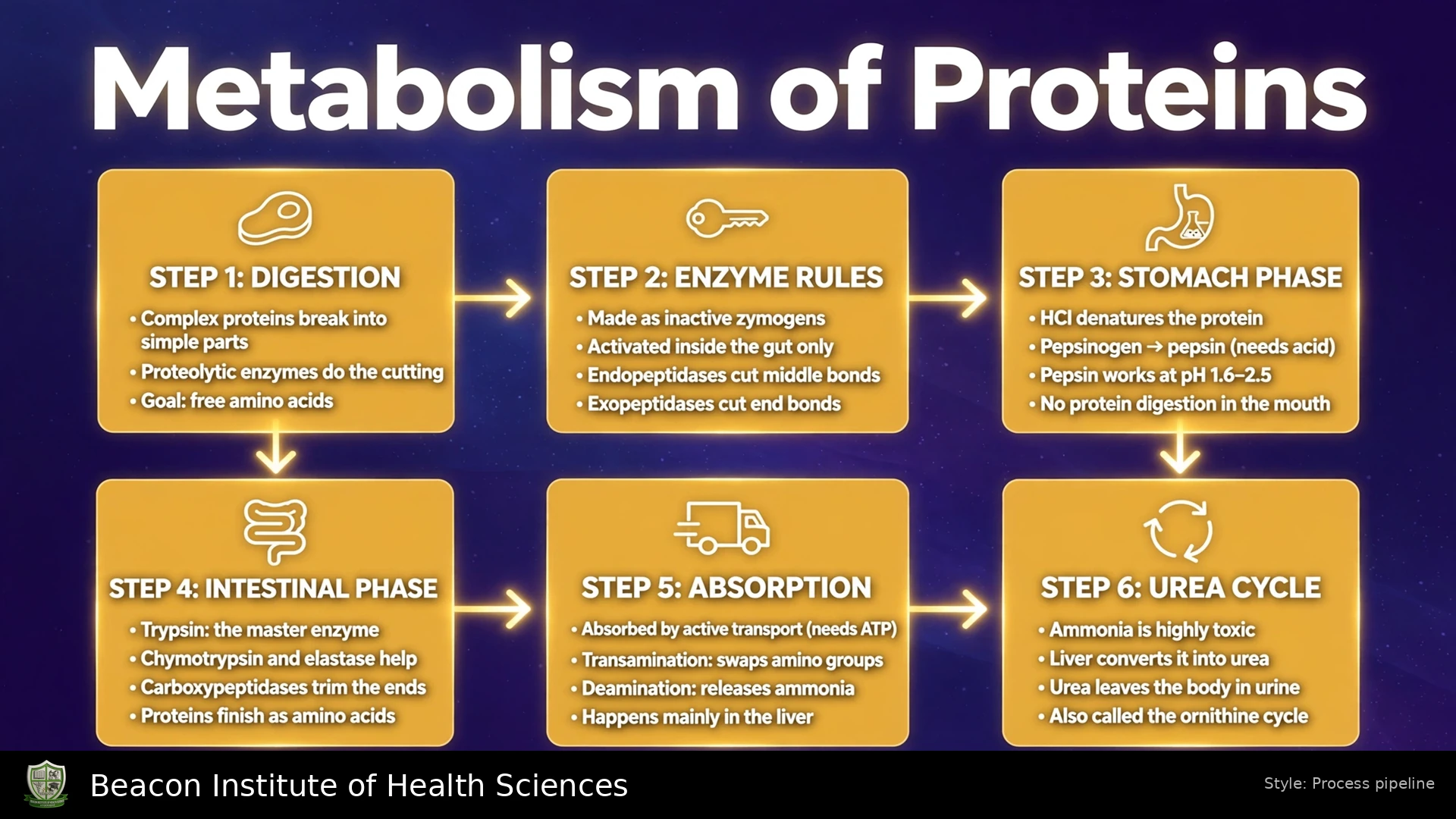
Protein digestion begins mainly in the stomach. Hydrochloric acid unfolds food proteins and provides the acidic environment needed for pepsin activity. Pepsin, released initially as pepsinogen, breaks peptide bonds and produces smaller polypeptides. Acid denaturation changes protein shape; it is different from enzymatic cleavage of peptide bonds.
In the small intestine, pancreatic bicarbonate helps neutralize acidic chyme. Pancreatic proteases are released as inactive precursors, reducing the risk of digesting the pancreas itself. Enteropeptidase activates trypsinogen to trypsin, which activates additional protease precursors. Trypsin, chymotrypsin, elastase and carboxypeptidases continue protein digestion.
Brush-border and intracellular peptidases complete digestion. Intestinal cells absorb free amino acids and small peptides through specialized transporters; absorbed peptides are largely broken into amino acids inside these cells. Amino acids then enter portal blood and reach the liver. Dietary proteins are therefore generally absorbed as their building blocks rather than as intact proteins.
Pathway: Dietary protein → smaller polypeptides → peptides and amino acids → intestinal absorption → portal blood → liver and other tissues.
3. The amino acid pool and nutritional classification
The amino acid pool consists of available free amino acids in blood and tissues. It receives amino acids from food, breakdown of existing body proteins and synthesis of nonessential amino acids. Its products support new proteins, nitrogen-containing compounds and energy metabolism. Unlike glycogen or triglyceride, protein has no dedicated storage depot that can be used without affecting functional tissue.
- Essential amino acids: Histidine, isoleucine, leucine, lysine, methionine, phenylalanine, threonine, tryptophan and valine must be supplied by the diet.
- Nonessential amino acids: The body can normally synthesize them. Examples include alanine, aspartate and glutamate. “Nonessential” does not mean biologically unimportant.
- Conditionally essential amino acids: In some growth or illness states, synthesis may not meet demand. The classification depends on the amino acid and clinical situation.
A varied diet can provide the required amino acids through animal or plant foods. Meeting protein needs also requires adequate total energy; otherwise more amino acids may be diverted from tissue maintenance to fuel production.
4. Protein synthesis and continuous turnover
Protein synthesis converts genetic information into an amino acid sequence. During transcription, information in DNA is copied into messenger RNA. During translation, a ribosome reads mRNA codons, and transfer RNA delivers corresponding amino acids. Peptide bonds join the growing chain until a stop signal terminates translation. Folding and further processing help produce a functional protein.
Information flow: DNA → mRNA → amino acid sequence → folded functional protein.
Synthesis is an energy-requiring anabolic process. It needs amino acids, cellular machinery and energy; eating protein does not cause an immediate gram-for-gram increase in muscle. Different tissues regulate synthesis according to growth, repair, hormones and disease.
Protein turnover is the continuous replacement of body proteins. Proteasomes and lysosomal pathways break down proteins, and many released amino acids are recycled. When breakdown exceeds synthesis over time, tissue protein is lost. Muscle wasting during severe illness reflects this imbalance, not simply a low dietary protein intake on one day.
5. Transamination: transferring amino nitrogen
Transamination transfers an amino group from an amino acid to a keto acid. A common acceptor is alpha-ketoglutarate, which becomes glutamate. The original amino acid becomes its corresponding keto acid. This reaction redistributes nitrogen without directly releasing free ammonia.
General reaction: Amino acid + alpha-ketoglutarate ⇌ corresponding alpha-keto acid + glutamate.
Aminotransferases use pyridoxal phosphate, derived from vitamin B6. Alanine aminotransferase (ALT) and aspartate aminotransferase (AST) are familiar examples. Their names describe the amino acids participating in the reactions.
- ALT: Alanine + alpha-ketoglutarate ⇌ pyruvate + glutamate.
- AST: Aspartate + alpha-ketoglutarate ⇌ oxaloacetate + glutamate.
These reversible reactions connect amino acid metabolism with carbohydrate intermediates and help synthesize some nonessential amino acids. Elevated blood ALT or AST can indicate cellular injury; these laboratory measurements do not directly measure a patient's dietary protein intake.
6. Deamination: releasing nitrogen for disposal
Deamination removes an amino group and can release ammonia or ammonium. Many amino acids first transfer nitrogen to glutamate. Mitochondrial glutamate dehydrogenase can then catalyze oxidative deamination, producing alpha-ketoglutarate and ammonium while reducing NAD+ or NADP+.
Simplified NAD+-linked reaction: Glutamate + NAD+ + water ⇌ alpha-ketoglutarate + NH4+ + NADH + H+.
The combination of transamination followed by glutamate deamination is often called transdeamination. It collects nitrogen from several amino acids into a common route. Do not assume every amino acid is directly deaminated by the same enzyme.
At physiological pH, much of this nitrogen is present as ammonium, NH4+. The term “ammonia” is commonly used clinically for the combined ammonia/ammonium system. Excess accumulation is harmful, so release of nitrogen must be coordinated with its transport and elimination.
7. Decarboxylation and specialized amino acid products
Decarboxylation removes a carboxyl group as carbon dioxide. It differs from transamination, which moves amino nitrogen, and from deamination, which removes amino nitrogen. Amino acid decarboxylation can generate biologically active amines.
Example: Glutamate → gamma-aminobutyric acid (GABA) + CO2, catalyzed by glutamate decarboxylase. Pyridoxal phosphate participates in this reaction.
This example shows that amino acids are precursors for signaling molecules as well as proteins and fuels. In an examination answer, identify the group removed and the product formed rather than treating all amino acid reactions as interchangeable.
8. Safe transport of nitrogen between organs
Peripheral tissues cannot simply release unlimited ammonia into the circulation. Glutamine and alanine provide important ways of carrying nitrogen to organs that can process it. Glutamine synthetase incorporates ammonium into glutamate to form glutamine, using ATP. Glutamine can travel to the liver or kidney, where its nitrogen is released through regulated reactions.
In muscle, transamination can transfer nitrogen to pyruvate, forming alanine. Alanine travels to the liver, where ALT regenerates pyruvate and transfers its nitrogen to glutamate. The liver can use pyruvate for glucose production and direct nitrogen toward urea formation. Returning glucose can support muscle energy needs; this relationship is the glucose-alanine cycle.
The kidneys also use glutamine in acid-base regulation. Ammonium excretion helps remove acid, particularly during acidosis. Therefore, urinary nitrogen disposal involves more than urea alone, even though urea is the major nitrogenous waste associated with amino acid breakdown.
9. The urea cycle: converting toxic nitrogen to urea
The complete urea cycle operates mainly in the liver. Its first two reactions occur in mitochondria, and the remaining reactions occur in the cytosol. Urea enters blood and is then removed mainly by the kidneys. One nitrogen in urea comes from free ammonia/ammonium and the other from aspartate; its carbon comes from bicarbonate.
- Carbamoyl phosphate formation: Carbamoyl phosphate synthetase I combines ammonia with bicarbonate, using two ATP. N-acetylglutamate activates this enzyme.
- Citrulline formation: Ornithine transcarbamylase transfers the carbamoyl group to ornithine. Citrulline is formed and moves to the cytosol.
- Argininosuccinate formation: Citrulline combines with aspartate through argininosuccinate synthetase. ATP is converted to AMP and pyrophosphate.
- Arginine formation: Argininosuccinate lyase splits argininosuccinate into arginine and fumarate. Fumarate connects the pathway with other metabolic reactions.
- Urea release: Arginase hydrolyzes arginine to urea and ornithine. Ornithine returns to the mitochondrion and is reused.
Energy accounting: Three ATP molecules are consumed per urea formed, equivalent to four high-energy phosphate bonds because one ATP is converted to AMP. The cycle disposes of nitrogen; it is not an ATP-generating pathway.
Worked example: For formation of 5 urea molecules, the direct cycle reactions consume 5 × 3 = 15 ATP molecules, equivalent to 5 × 4 = 20 high-energy phosphate bonds. This calculation concerns the cycle's direct cost, not the net energy of all linked metabolic pathways.
Impaired hepatic processing or an inherited enzyme defect can lead to hyperammonemia. Urea and uric acid are different compounds: urea is a major product of amino nitrogen disposal, whereas uric acid is associated with purine breakdown.
10. Carbon skeletons: glucogenic and ketogenic routes
After nitrogen is handled, amino acid carbon skeletons enter metabolism as pyruvate, acetyl-CoA, acetoacetate or citric acid cycle intermediates such as alpha-ketoglutarate, succinyl-CoA, fumarate and oxaloacetate. Their entry points determine their possible metabolic products.
- Glucogenic amino acids: Produce substrates that can contribute to net glucose synthesis. Alanine produces pyruvate; aspartate produces oxaloacetate.
- Ketogenic amino acids: Produce acetyl-CoA or acetoacetate. Leucine and lysine are exclusively ketogenic.
- Both glucogenic and ketogenic: Some amino acids provide products in both categories. Isoleucine is an example.
Acetyl-CoA cannot provide net glucose production in humans. Therefore, being usable for energy does not automatically mean an amino acid is glucogenic. Carbon skeletons can also be oxidized through the citric acid cycle and oxidative phosphorylation or contribute to lipid synthesis when conditions favor it.
11. Nitrogen balance and a worked calculation
Nitrogen balance compares nitrogen intake with total nitrogen loss over the same period. Positive balance occurs when intake exceeds loss, as may happen during growth or recovery. Negative balance means loss exceeds intake and may accompany inadequate intake, trauma or severe illness. Approximate balance is expected in a stable adult maintaining body protein.
Formula: Nitrogen balance = nitrogen intake − total nitrogen losses.
For a teaching estimate, dietary protein contains about 16% nitrogen: nitrogen intake in grams ≈ protein intake in grams ÷ 6.25. The exact factor varies with protein composition. Total loss includes urinary and nonurinary nitrogen; urinary urea nitrogen alone is not the total.
Worked example: Assume an adult consumes 87.5 g protein in 24 hours and a complete assessment estimates total nitrogen losses at 16 g over that same period. Nitrogen intake = 87.5 ÷ 6.25 = 14 g. Balance = 14 − 16 = −2 g nitrogen per day, indicating negative balance under these assumptions.
This is an educational calculation, not a feeding prescription. Incomplete collections, changing renal function, dialysis and large wound or gastrointestinal losses can affect interpretation. A nutrition team must consider the clinical setting and measurement quality.
12. Energy use in fed, fasting and illness states
After a meal, amino acids support protein synthesis and other biosynthetic needs. Surplus amino acids are not retained in a separate protein store: their nitrogen is disposed of and their carbon skeletons enter metabolic pathways. Adequate carbohydrate and fat energy can help spare amino acids for their structural and functional roles.
During fasting, body protein breakdown can supply amino acids for gluconeogenesis and energy. In prolonged fasting, greater ketone use helps reduce, but does not eliminate, the need to break down protein. Stress hormones and inflammation during major illness can increase catabolism even when food is being provided.
Worked energy example: Using the conventional dietary estimate of 4 kcal per gram, 25 g of protein contributes approximately 25 × 4 = 100 kcal. This food-energy estimate is not the ATP yield of one amino acid. ATP yield varies with its carbon skeleton and the reactions required for nitrogen handling.
These mechanisms explain why nutrition assessment considers both energy and protein. Simply adding protein without addressing inadequate intake, feeding intolerance or the underlying illness may not correct tissue loss.
13. Clinical interpretation: liver, kidney and nutrition markers
Blood urea nitrogen (BUN) measures the nitrogen component of urea in blood. A rise can reflect reduced kidney clearance, dehydration or increased protein breakdown, among other causes. A low value can occur with low intake or impaired urea production. Interpret BUN with creatinine, hydration, symptoms and the wider clinical assessment; it is not a stand-alone nutrition test.
Ammonia testing may be used when a disorder of nitrogen disposal is suspected. New confusion, marked drowsiness, vomiting or seizures require prompt clinical assessment; these findings are not specific to hyperammonemia. Follow local specimen-handling requirements because collection and processing can affect the result.
Albumin and prealbumin are affected by inflammation and other disease-related processes. Their concentrations should not be used alone to diagnose malnutrition or estimate total muscle protein. Review intake, weight history, muscle and fat loss, functional status and clinical findings through the appropriate nutrition assessment process.
Inherited amino acid disorders are another clinical connection. For example, impaired phenylalanine metabolism in phenylketonuria requires specialist management. Urea cycle disorders may cause dangerous ammonia accumulation. Do not translate a general lecture into unsupervised dietary restriction or treatment.
14. Nursing applications and patient scenarios
Scenario 1 - Poor intake after surgery: A patient eats little for several days and is losing strength. Explain that inadequate energy and protein can shift turnover toward tissue loss. Record actual intake, assess barriers such as nausea or swallowing difficulty, monitor the clinical course and refer for nutrition review. Do not rely on a single albumin result to describe nutritional status.
Scenario 2 - Kidney disease and dialysis: A patient asks whether all kidney patients should avoid protein. Explain that requirements vary with kidney function, treatment and nutritional state. Dialysis can remove amino acids and protein-related nutrients, so needs differ from those before dialysis. Reinforce the individualized renal dietitian's plan rather than giving blanket protein restriction advice.
Scenario 3 - Liver disease with new confusion: Recognize altered mental status promptly and escalate assessment. Link impaired nitrogen handling to possible ammonia accumulation, while remembering other causes of confusion. Follow prescribed monitoring and treatment. Dietary changes require the clinical team's plan; do not automatically remove all protein.
Scenario 4 - A slowly healing wound: Review intake, recent weight change and feeding tolerance alongside the wound assessment. Proteins supply amino acids for tissue repair, but healing also depends on adequate energy, perfusion, infection control and other nutrients. Coordinate care with the wound and nutrition teams and document progress.
- Monitor nutritional intake and relevant fluid balance consistently.
- Observe strength, mobility and changes in body weight in their clinical context.
- Check prescribed feeding plans, tolerance and appropriate laboratory trends.
- Explain food choices clearly and avoid suggesting that one food or supplement cures metabolic disease.
15. Revision questions and key distinctions
- What starts protein digestion? Gastric acid and pepsin begin the main digestive process; pancreatic and intestinal enzymes continue it.
- Does transamination release free ammonia? No. It transfers an amino group; deamination can subsequently release ammonium.
- What does decarboxylation remove? A carboxyl group as carbon dioxide.
- Where is urea formed and removed? The complete cycle is mainly hepatic; renal excretion removes urea from the circulation.
- Why are three ATP and four high-energy bonds both correct? One of the three ATP molecules is converted to AMP rather than ADP.
- Which amino acids are exclusively ketogenic? Leucine and lysine.
- What does negative nitrogen balance mean? Estimated total nitrogen loss exceeds intake during the assessment period.
- Can albumin alone diagnose malnutrition? No. It is influenced by inflammation and requires wider clinical interpretation.
Follow the overall sequence when revising: protein digestion and absorption → amino acid pool → synthesis or breakdown → safe nitrogen disposal plus carbon-skeleton metabolism. Keep the amino group and carbon skeleton separate in your explanation, then connect the pathways to patient assessment.