
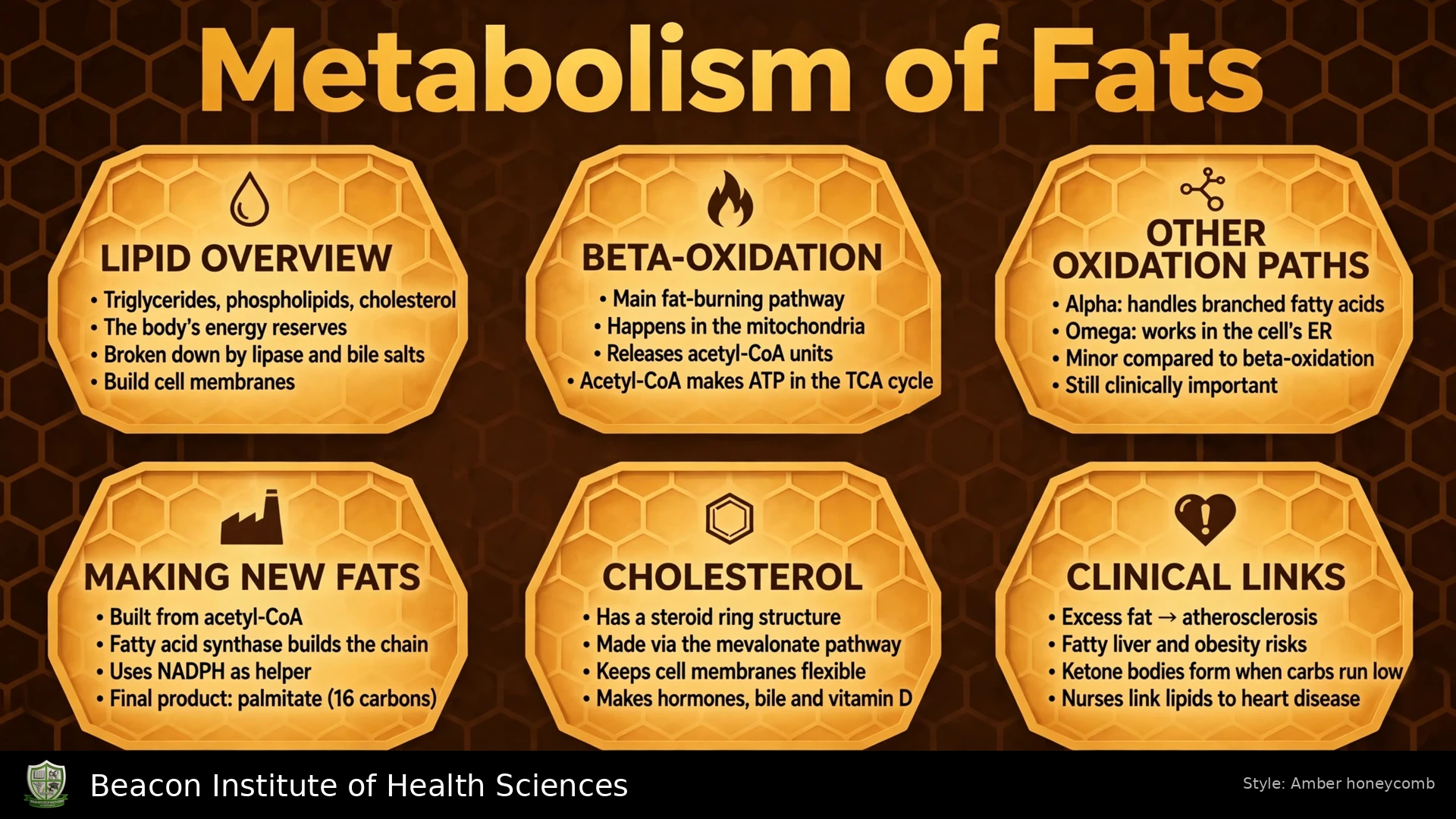
1. What fat metabolism means
Fat metabolism includes the digestion, absorption, transport, storage, synthesis and breakdown of lipids. These processes allow the body to obtain energy from dietary and stored fat while supplying molecules for membranes, signaling and other functions. Triacylglycerol, also called triglyceride, is the main storage form of fat and consists of glycerol joined to three fatty acids.
Fat provides approximately 9 kcal per gram, compared with approximately 4 kcal per gram from carbohydrate or protein. This nutritional value is different from the number of ATP molecules produced by oxidation of one fatty acid. Nurses need both concepts when interpreting nutrition and metabolic changes.
- Lipolysis releases fatty acids and glycerol from triglycerides.
- Beta-oxidation breaks fatty acyl-CoA into smaller units, mainly acetyl-CoA.
- Lipogenesis builds fatty acids and stores energy as triglycerides.
- Ketogenesis produces ketone bodies from acetyl-CoA in the liver.
2. Digestion of dietary fats
Most dietary lipid digestion occurs in the small intestine. Bile salts help disperse large fat droplets, increasing the surface available to digestive enzymes. This emulsification is a physical process; bile is not itself a lipase. Pancreatic lipase, assisted by colipase, hydrolyzes triglycerides mainly into free fatty acids and monoacylglycerols.
Digestion products combine with bile salts in mixed micelles, which help deliver poorly water-soluble lipids to the intestinal surface. Micelles are different from chylomicrons: micelles participate in intestinal absorption, whereas chylomicrons are transport particles assembled by intestinal cells.
Reduced bile delivery, pancreatic enzyme deficiency or intestinal disease can impair fat absorption. Nursing observations may include greasy stools, weight change and nutritional concerns. These findings require assessment rather than diagnosis from appearance alone.
3. Absorption and chylomicron transport
After entering intestinal cells, most long-chain fatty acids and monoacylglycerols are reassembled into triglycerides. They are packaged with cholesterol, phospholipids and proteins into chylomicrons. Chylomicrons enter intestinal lymph and subsequently the bloodstream. Shorter-chain fatty acids can reach portal blood more directly.
In capillaries of tissues such as adipose tissue and muscle, lipoprotein lipase helps release fatty acids from circulating triglycerides. These fatty acids can be stored or used for energy. Chylomicron remnants are taken up by the liver. Lipoprotein lipase at capillary surfaces must not be confused with enzymes that mobilize fat already stored within adipocytes.
4. Lipoproteins and circulation
Lipids require transport systems because they do not dissolve freely in the watery plasma. Lipoproteins contain both lipid and protein components. Their roles differ, and their names do not describe a single kind of dietary fat.
- Chylomicrons mainly transport dietary triglyceride from the intestine.
- VLDL carries triglyceride produced or packaged by the liver to other tissues.
- LDL participates in cholesterol delivery to cells.
- HDL participates in cholesterol transport, including movement toward hepatic handling.
- Non-esterified fatty acids released from adipose tissue circulate largely bound to albumin.
A lipid profile measures selected circulating lipid concentrations. It does not directly measure the rate of beta-oxidation. Interpretation requires the person's clinical context and cardiovascular risk assessment; one value should not be treated as a complete metabolic diagnosis.
5. Storage and mobilization of triglycerides
Adipose tissue stores triglycerides in lipid droplets. After a meal, insulin supports energy storage and suppresses excessive mobilization of stored fat. When energy intake falls or energy demand increases, stored triglycerides can be hydrolyzed through coordinated actions of adipose triglyceride lipase, hormone-sensitive lipase and monoacylglycerol lipase.
Triglyceride + 3 water molecules → glycerol + 3 fatty acids
Catecholamine signaling can stimulate adipose lipolysis during stress or activity. Released fatty acids travel to tissues for oxidation, while glycerol can travel to the liver for further metabolism. The balance between storage and release depends on hormonal signals, substrate availability and tissue requirements, not simply the time since the last meal.
6. Fatty acid activation and mitochondrial entry
Before oxidation, a fatty acid is activated to fatty acyl-CoA by an acyl-CoA synthetase. ATP is converted to AMP and pyrophosphate. Because pyrophosphate is subsequently hydrolyzed, activation costs two high-energy phosphate equivalents, even though only one ATP molecule is used in the initial reaction.
Fatty acid + CoA + ATP → fatty acyl-CoA + AMP + pyrophosphate
Long-chain fatty acyl groups use the carnitine shuttle to cross the inner mitochondrial membrane. CPT I forms acylcarnitine, a translocase carries it across, and CPT II restores fatty acyl-CoA on the matrix side. The shuttle transfers the acyl group; it does not carry cytosolic CoA intact into the matrix. Shorter-chain fatty acids have different transport requirements.
7. The four reactions of beta-oxidation
The principal oxidation of ordinary long-chain fatty acids takes place in the mitochondrial matrix. Each cycle acts near the beta carbon and shortens the fatty acyl chain by two carbons. The pathway repeats until the chain has been broken into acetyl-CoA units.
- First oxidation: acyl-CoA dehydrogenase introduces a double bond and transfers reducing equivalents through a flavin-linked system.
- Hydration: enoyl-CoA hydratase adds water across the double bond.
- Second oxidation: hydroxyacyl-CoA dehydrogenase produces a keto group and NADH.
- Thiolysis: thiolase uses CoA to split off acetyl-CoA, leaving a chain two carbons shorter.
For a typical saturated even-chain fatty acid, each cycle yields one NADH and one FADH2-equivalent, along with chain shortening. The final cycle of a four-carbon chain gives two acetyl-CoA molecules. Beta-oxidation itself does not directly produce large amounts of ATP; its products support subsequent ATP production.
8. Linking oxidation to ATP production
Acetyl-CoA can enter the citric acid cycle, where further oxidation produces carbon dioxide and reduced electron carriers. NADH and flavin-linked reducing equivalents support electron transport and oxidative phosphorylation. Oxygen is the terminal electron acceptor, so sustained fatty acid oxidation depends on functioning aerobic energy metabolism.
Using common modern estimates, one NADH corresponds to approximately 2.5 ATP and one FADH2-equivalent to approximately 1.5 ATP. One acetyl-CoA oxidized in the citric acid cycle contributes approximately 10 ATP equivalents. These are accounting estimates, not fixed measurements in every cell.
Fatty acids are therefore energy-rich fuels, but they cannot replace every role of glucose. Red blood cells lack mitochondria and cannot obtain ATP through fatty acid beta-oxidation. This is one reason blood glucose must remain available even when fat stores are abundant.
9. Worked example: palmitate oxidation
Palmitate is a saturated fatty acid containing 16 carbons. For a saturated even-chain fatty acid with n carbons, the number of acetyl-CoA units is n/2, and the number of beta-oxidation cycles is n/2 - 1.
- Acetyl-CoA units: 16/2 = 8.
- Beta-oxidation cycles: 16/2 - 1 = 7.
- Products from seven cycles: 7 NADH and 7 FADH2-equivalents.
- ATP equivalents from these carriers: (7 × 2.5) + (7 × 1.5) = 28.
- ATP equivalents from eight acetyl-CoA: 8 × 10 = 80.
- Total before activation: 28 + 80 = 108.
- Subtract activation cost: 108 - 2 = approximately 106 net ATP.
This calculation assumes complete mitochondrial oxidation and the stated modern carrier yields. Older teaching conventions use different ATP-per-carrier values and may produce different totals. State the convention rather than combining old and new numbers. The 106 estimate applies to palmitate, not every fat molecule.
10. Other fatty acids and oxidation pathways
Unsaturated fatty acids require additional enzymes to handle existing double bonds; their ATP accounting differs from the simple saturated-chain example. Odd-chain fatty acids leave a final three-carbon propionyl-CoA unit, which can be converted to succinyl-CoA through reactions involving biotin and vitamin B12.
Very-long-chain fatty acids initially undergo shortening in peroxisomes. Peroxisomal oxidation has different energy coupling from mitochondrial beta-oxidation and should not be assigned the same ATP yield automatically. Other specialized pathways help process particular fatty acid structures. For introductory revision, master ordinary mitochondrial beta-oxidation first, then recognize these exceptions.
11. Ketogenesis in the liver
When hepatic fatty acid oxidation supplies substantial acetyl-CoA, some acetyl-CoA can be used to form ketone bodies in liver mitochondria. This becomes especially important during fasting and marked insulin deficiency. Increased gluconeogenesis and changes in oxaloacetate availability can also favor diversion of acetyl-CoA toward ketogenesis.
- Acetoacetate is a metabolically useful ketone body.
- Beta-hydroxybutyrate is conventionally grouped with ketone bodies, although chemically it is a hydroxy acid.
- Acetone forms from acetoacetate and can be exhaled.
Ketone bodies are water-soluble fuels that can travel to other tissues. Ketogenesis is a controlled physiological pathway, but excessive production in some clinical conditions can contribute to dangerous acidosis. The presence of ketones alone does not establish diabetic ketoacidosis.
12. Ketone use and ketoacidosis
Extrahepatic tissues with mitochondria can convert acetoacetate or beta-hydroxybutyrate into acetyl-CoA for energy production. The liver makes ketone bodies but lacks the SCOT enzyme needed for their principal utilization pathway. Red blood cells cannot oxidize ketone bodies because they lack mitochondria. During prolonged fasting, the brain increases its use of ketones, reducing but not eliminating glucose requirements.
Diabetic ketoacidosis involves excessive ketone accumulation with metabolic acidosis in the setting of diabetes-related insulin deficiency. It can occur without extremely high glucose, including in some patients taking SGLT2 inhibitors. Physiological fasting ketosis and DKA must not be treated as equivalent.
Nursing connection: vomiting, abdominal pain, dehydration, deep rapid breathing or altered consciousness in a person at risk require prompt assessment and escalation. Follow the local emergency pathway for glucose, ketone and acid-base assessment. Treatment requires prescribed fluid, insulin and electrolyte management with monitoring; do not infer a treatment dose from this lecture.
13. Fatty acid synthesis and its regulation
Fatty acid synthesis mainly occurs in the cytosol, particularly in tissues such as liver and adipose tissue. Carbon from mitochondrial acetyl-CoA is transferred through citrate-based processes. Acetyl-CoA carboxylase forms malonyl-CoA, using ATP and biotin. Fatty acid synthase uses carbon units and NADPH to build the chain.
Synthesis and oxidation are not simply one pathway running in opposite directions. They use different locations, enzymes and electron carriers. NADPH supports synthesis, whereas NADH and flavin-linked carriers produced during oxidation support energy transfer.
Malonyl-CoA inhibits CPT I and thereby limits long-chain fatty acid entry for oxidation when synthesis is active. Insulin generally favors storage and synthesis after a meal. Fasting and increased energy demand favor mobilization and oxidation. Coordinated regulation reduces wasteful simultaneous synthesis and breakdown.
14. Glycerol, cholesterol and metabolic integration
Glycerol can be converted to glycerol-3-phosphate and then to a glycolytic intermediate, linking triglyceride breakdown with carbohydrate pathways. In the liver it can contribute to gluconeogenesis. In contrast, acetyl-CoA from ordinary even-chain fatty acids does not yield net glucose in humans. Odd-chain fatty acid carbon has a different metabolic fate through propionyl-CoA.
Cholesterol metabolism is related to, but distinct from, triglyceride energy metabolism. Cholesterol contributes to membranes and is a precursor for steroid hormones and bile acids. It is not oxidized by beta-oxidation like a fatty acid. The liver regulates its synthesis, uptake and elimination pathways.
A balanced account of lipid metabolism should therefore separate energy storage, fatty acid oxidation, ketone use and cholesterol transport. The broad label “fat” does not make these processes interchangeable.
15. Nursing scenarios and revision
Scenario: reduced intake over several days. A patient has eaten poorly because of illness. Fat mobilization may increase, but substantial fat stores do not rule out malnutrition or other nutritional deficiencies. Assess intake, weight trends, hydration, symptoms and the prescribed nutrition plan; communicate concerns to the clinical team.
Scenario: greasy stools and weight loss. A patient reports persistent bulky, greasy stools. Consider the need for clinical evaluation of malabsorption and record symptoms and dietary history. Do not simply recommend complete fat avoidance: energy and essential nutrient needs still require assessment.
Scenario: diabetes, vomiting and deep breathing. Recognize possible metabolic deterioration, escalate promptly and follow the emergency assessment pathway. A moderately elevated glucose reading does not safely exclude DKA in every setting. Review relevant medicines and communicate observations without delaying assessment.
- Explain how dietary fat reaches muscle and adipose tissue.
- Distinguish lipoprotein lipase from stored-fat mobilization enzymes.
- Name the four beta-oxidation reactions in order.
- Calculate palmitate cycles, acetyl-CoA and net ATP using explicit assumptions.
- Explain why the liver produces ketones but does not use them through the principal ketolysis pathway.
- Distinguish fasting ketosis from a clinical ketoacidotic emergency.
- Compare fatty acid synthesis with oxidation and explain the role of malonyl-CoA.
Self-check: trace a dietary triglyceride from intestinal digestion to tissue storage, then trace a stored fatty acid from mobilization to mitochondrial ATP production. Connect each stage with one relevant nursing observation or assessment.