
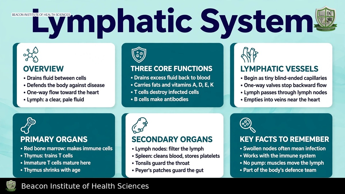
Reference-based BSN study notes for Anatomy and Physiology I.
Learning objectives and functions
Describe lymph formation and return, identify lymphatic vessels and major ducts, compare lymphoid organs, and connect fluid balance, fat absorption and immune surveillance. This unit focuses on Anatomy and Physiology I structure and function. It complements microbiology's body-defence topic but adds drainage pathways and organ anatomy rather than duplicating an immunity summary.
Formation of lymph
Blood capillaries exchange water and solutes with interstitial spaces. Net filtered fluid and escaped proteins require lymphatic return. When interstitial fluid enters a lymphatic vessel it is called lymph. Its composition varies by region and reflects tissue fluid; it can contain proteins, immune cells and absorbed intestinal lipids. Lymph is not a separate type of blood and normally contains far fewer erythrocytes. Return of tissue fluid and proteins helps maintain blood volume and prevent interstitial accumulation.
Capillaries and collecting vessels
Initial lymphatic capillaries begin as blind-ended, highly permeable channels. Overlapping endothelial arrangements and supporting filaments facilitate entry when tissue pressure increases. These join collecting vessels with valves that favour forward flow. Lymph passes through regional nodes before reaching larger trunks and ducts, although precise routes vary by region.
Unlike systemic circulation, lymph flow has no central heart-like pump. Skeletal-muscle movement, breathing-related pressure changes, vessel-wall contraction and arterial pulsation help move it. Valves limit reflux. Impaired movement or obstruction can reduce effective drainage, but oedema may also arise from cardiac, renal, hepatic, venous or other causes. Do not diagnose lymphatic obstruction solely from swelling.
Major ducts and return to veins
The right lymphatic duct typically drains the right side of the head and neck, right upper limb and right thorax into the right venous angle near the junction of internal jugular and subclavian veins. The thoracic duct drains most of the remaining body and enters near the corresponding left venous angle. Its lower origin is often associated with the cisterna chyli, which receives intestinal and lumbar drainage, with anatomical variation.
These drainage territories are asymmetric: the right duct does not drain the whole right half of the body. Learning this distinction avoids a common examination error. Superficial and deep collecting routes contribute to regional drainage, with communication between pathways.
Lymph nodes
Nodes are small encapsulated organs positioned along lymphatic vessels. Afferent vessels bring lymph in; efferent vessels carry it out at the hilum. Lymph passes through sinuses containing cells that support filtration and immune surveillance. The cortex contains follicles rich in B cells; the paracortex contains many T cells; medullary regions contain cords and sinuses. Nodes facilitate interaction among antigens, antigen-presenting cells and lymphocytes.
Groups include cervical, axillary, mediastinal, abdominal, pelvic and inguinal nodes. Enlargement can reflect infection, inflammation or malignancy and does not by itself establish a cause. Clinical assessment considers location, tenderness, consistency, distribution, duration and the wider history. Do not confuse lymph nodes with endocrine glands simply because both may be called glands in everyday language.
Primary and secondary lymphoid organs
Primary lymphoid organs support lymphocyte development: bone marrow is central to blood-cell production and B-cell maturation, while the thymus supports T-cell maturation and selection. The thymus is prominent in childhood and undergoes age-related involution, but immune function does not simply cease when it becomes smaller.
Secondary lymphoid organs include lymph nodes, spleen and mucosa-associated lymphoid tissues. The spleen lies in the left upper abdomen; white pulp supports immune responses and red pulp filters blood and removes ageing or damaged erythrocytes. Unlike lymph nodes, the spleen principally filters blood rather than lymph. Its vascular structure makes injury clinically significant.
Tonsils and other mucosal lymphoid tissues encounter antigens at entry surfaces. Peyer's patches in the ileum and lymphoid tissue associated with the appendix contribute to intestinal immune surveillance. These tissues cooperate with epithelial barriers and other defence mechanisms; they do not replace those barriers.
Intestinal lipid absorption
Lacteals are specialised lymphatic capillaries in small-intestinal villi. Many absorbed dietary long-chain lipids are packaged into chylomicrons, which enter lacteals and travel through lymph before joining venous blood. Lipid-rich intestinal lymph is called chyle. This pathway differs from the direct portal-blood transport of many water-soluble nutrients. Not every dietary lipid follows the identical route, so avoid stating that all absorbed fat bypasses portal blood without qualification.
Integration and nursing relevance
Lymphatic and cardiovascular systems cooperate in fluid balance; lymphoid tissues cooperate with innate and adaptive immunity. Adaptive responses involve antigen-specific lymphocytes and memory, while innate defences respond through more general mechanisms. Anatomy explains the route and sites of these interactions, rather than supplying a complete immunology treatment course.
Observe and document swelling, skin integrity and relevant node findings within scope. Following surgery or node removal, lymphatic drainage may be altered and patient-specific precautions matter. New unilateral swelling, redness, pain or systemic symptoms require assessment and escalation; do not begin massage or compression based only on a classroom explanation. Protection from skin injury and infection can be relevant in patients with impaired drainage, governed by their care plan.
Revision checks
Trace fluid from tissue space to initial capillary, collecting vessel, node, trunk, duct and venous circulation. Contrast the right duct's territory with the thoracic duct's. Compare node filtration with splenic filtration; identify where B and T cells mature; and explain why lacteals are important. Distinguish fluid-return function from immune function while recognising that the same system supports both.