
1. Meaning and importance of enzymology
Enzymology is the study of enzymes: their structure, catalytic action, classification, reaction rates, regulation and applications in health and disease. An enzyme is a biological catalyst that increases the rate of a chemical reaction without being consumed in the overall reaction. Most enzymes are proteins. Some RNA molecules also catalyze reactions and are called ribozymes; the ribosome's peptide-bond-forming activity is an important example.
Human cells depend on enzymes for digestion, ATP production, synthesis and breakdown of biomolecules, DNA replication, blood coagulation, detoxification and communication. A reaction that is chemically possible may proceed too slowly to sustain life unless an enzyme accelerates it. Enzymes allow these processes to occur under conditions compatible with living tissues.
For nursing students, enzymology connects biochemistry with laboratory interpretation, nutrition, inherited disorders, medication action and patient monitoring. A raised serum enzyme activity may indicate tissue injury, while a deficiency of intracellular enzyme activity may impair a metabolic pathway. These are different situations and should not be confused.
2. Essential terms and properties
- Substrate: The reactant on which an enzyme acts. A reaction may require one or several substrates.
- Product: A molecule formed by the enzyme-catalyzed reaction.
- Active site: The three-dimensional region that binds the substrate and contains groups involved in catalysis.
- Enzyme-substrate complex: The temporary association of enzyme and substrate during the catalytic cycle.
- Catalysis: Acceleration of a reaction by providing a pathway with a lower activation-energy barrier.
- Specificity: Preference for particular substrates, chemical bonds or stereochemical forms.
- Enzyme activity: The rate of a defined reaction measured under specified conditions.
Enzymes are highly effective and often act at low concentrations. The enzyme is regenerated at the end of a catalytic cycle, although enzymes can be damaged, inhibited or degraded over time. Protein enzymes require an appropriate folded structure. Disruption of that structure may reduce activity even when peptide bonds remain intact.
Enzymes do not supply unlimited energy, change the equilibrium constant or change the overall free-energy difference between reactants and products. They accelerate the approach to equilibrium. For a reversible reaction, the catalyst facilitates both directions. Cells can drive an unfavorable process by coupling it to a favorable reaction, such as ATP hydrolysis; the energy comes from the coupled reaction, not from the enzyme itself.
3. Enzyme structure and substrate specificity
An active site may contain amino acids that are far apart in the primary sequence but become neighbors when the protein folds. Binding depends on shape, charge, hydrogen bonding and hydrophobic interactions. The binding region recognizes substrates, while catalytic groups facilitate their conversion. Many enzymes contain several subunits, and some have separate regulatory sites.
Absolute specificity means that an enzyme strongly favors one substrate, as illustrated by urease acting on urea. Group specificity means that related substrates containing a particular chemical group may be accepted. Bond specificity means that a certain bond type is targeted, as with proteases hydrolyzing peptide bonds. Stereospecificity means discrimination between spatial arrangements; many metabolic enzymes accept one stereoisomer much more readily than another.
Specificity is not an absolute rule that every enzyme has only one possible substrate. Hexokinase, for example, can phosphorylate several hexoses. Its physiological role depends on substrate availability, cellular location and the properties of the enzyme.
Lock-and-key and induced-fit models
The lock-and-key model describes a substrate fitting a complementary active site and is useful for introducing specificity. It oversimplifies enzymes as rigid structures. The induced-fit model recognizes that substrate binding can change enzyme conformation, position catalytic groups and help stabilize the transition state. Both enzyme and substrate can undergo changes during binding.
4. Mechanism of enzyme action
A simplified catalytic sequence is:
E + S <=> ES -> E + P
Here E is enzyme, S is substrate, ES is the enzyme-substrate complex and P is product. Actual reactions may involve multiple substrates, intermediates and product-release steps.
- The substrate encounters the enzyme and binds in a suitable orientation.
- Binding forms an enzyme-substrate complex and may cause conformational adjustment.
- Catalytic groups stabilize the transition state and support bond breaking or bond formation.
- Products form and are released.
- The regenerated enzyme can begin another cycle.
Activation energy is the barrier that reactants must overcome to reach a transition state. Lowering this barrier increases the fraction of molecular encounters that lead to reaction. An enzyme may bring reactants close together, orient them correctly, transfer protons, form a temporary covalent intermediate or use a metal ion to stabilize charge. These are complementary catalytic mechanisms rather than mutually exclusive categories.
In acid-base catalysis, groups donate or accept protons. In covalent catalysis, a temporary bond forms between enzyme and substrate and is subsequently broken. In metal-ion catalysis, a metal can assist substrate binding, electron transfer or charge stabilization. An active site often combines several mechanisms.
5. Cofactors, coenzymes and holoenzymes
Some enzymes function independently; others require a non-protein component. A cofactor is a required non-protein helper and can be an inorganic ion or an organic molecule. An organic cofactor is a coenzyme. Some introductory classifications use "cofactor" specifically for inorganic ions, but the broader definition includes coenzymes.
An apoenzyme is the protein component without its required cofactor. The complete functional enzyme-cofactor assembly is a holoenzyme. A prosthetic group is tightly associated with an enzyme, sometimes through a covalent bond. A cosubstrate binds, is chemically altered during the reaction and is regenerated through another reaction.
Metal examples include magnesium in many ATP-dependent reactions, zinc in carbonic anhydrase and iron in heme-containing enzymes. Cofactor requirements are enzyme-specific; one metal should not be assumed to substitute for another.
Important vitamin-derived coenzymes include:
- Thiamine, vitamin B1: thiamine pyrophosphate in oxidative decarboxylation and transketolase reactions.
- Riboflavin, vitamin B2: FAD and FMN in oxidation-reduction reactions.
- Niacin, vitamin B3: NAD and NADP in electron-transfer reactions.
- Pantothenic acid, vitamin B5: coenzyme A in acyl-group transfer.
- Pyridoxine and related vitamin B6 forms: pyridoxal phosphate in many amino-acid reactions.
- Biotin, vitamin B7: a bound coenzyme in carboxylation reactions.
- Folate, vitamin B9: tetrahydrofolate derivatives in one-carbon transfer.
- Vitamin B12: coenzyme forms required by methionine synthase and methylmalonyl-CoA mutase.
Vitamin deficiency can impair enzyme-dependent pathways even when the enzyme protein is present. Nutritional assessment therefore has biochemical importance. Vitamin supplementation does not mean all enzyme reactions become faster; benefit depends on deficiency, absorption, clinical indication and the specific pathway.
6. Enzyme naming and classification
Many enzyme names end in "-ase" and describe a substrate or reaction: lactase, lipase and alcohol dehydrogenase. Historical names such as pepsin and trypsin do not follow that pattern. An Enzyme Commission, or EC, number classifies an enzyme by the reaction it catalyzes. The four parts indicate class, subclass, sub-subclass and an identifying number. It is a reaction classification, not a unique label for a particular gene or protein sequence.
The current IUBMB system has seven major classes. Older notes may list six because translocases were added later.
- Oxidoreductases, EC 1: Catalyze oxidation-reduction. Lactate dehydrogenase interconverts lactate and pyruvate with NAD/NADH participation.
- Transferases, EC 2: Transfer a group between molecules. Aminotransferases transfer amino groups; hexokinase transfers a phosphate from ATP to a hexose.
- Hydrolases, EC 3: Cleave bonds using water. Examples include lipases, proteases, phosphatases and many digestive enzymes.
- Lyases, EC 4: Remove or add groups by mechanisms other than hydrolysis or oxidation, often forming or using a double bond. Aldolase and many decarboxylases are examples.
- Isomerases, EC 5: Rearrange atoms within a molecule. Phosphoglucose isomerase interconverts glucose-6-phosphate and fructose-6-phosphate.
- Ligases, EC 6: Join molecules using energy coupled to nucleoside-triphosphate cleavage or a comparable energy source. DNA ligase forms phosphodiester bonds during DNA processing.
- Translocases, EC 7: Catalyze movement of ions or molecules across membranes, or their separation within membranes, coupled to a driving reaction. ATP-dependent ion pumps are examples.
Do not classify an enzyme only from a familiar name. Consult the reaction and current EC assignment when a precise classification is required.
7. Factors affecting enzyme activity
Temperature and pH
Increasing temperature often increases reaction rate over a limited range because molecular movement increases. At higher temperatures, structural instability and denaturation may reduce activity. The measured optimum depends on the enzyme, assay duration and experimental conditions. Many human enzymes operate effectively near physiological temperature, but 37 degrees Celsius is not a universal optimum for every enzyme. Cooling usually slows reactions without necessarily destroying the enzyme.
pH changes the ionization of substrates and active-site groups. This can alter binding and catalysis; extreme pH may also disrupt folding. Pepsin functions in an acidic gastric environment, while pancreatic digestive enzymes act in the less acidic small intestine. Each enzyme has a characteristic activity-pH relationship. Activity outside the preferred range can fall before irreversible denaturation occurs.
Enzyme and substrate concentrations
When sufficient substrate is available and conditions are fixed, increasing the concentration of active enzyme usually increases the initial reaction rate proportionally. If substrate becomes limiting, that relationship cannot be assumed.
At a fixed enzyme concentration, increasing substrate initially increases reaction rate. Many enzymes approach a maximum rate as active sites become occupied. Substrate saturation is not enzyme destruction: additional substrate simply produces little further increase in rate under those conditions. Some enzymes show substrate inhibition at very high concentrations or exhibit cooperative behavior rather than a simple saturation curve.
Additional influences
Required cofactors, ionic strength, inhibitors, activators, product accumulation and substrate accessibility also influence measured activity. In living cells, enzyme location and transport of substrates between compartments can matter as much as enzyme quantity. A laboratory value must therefore specify the reaction and measurement conditions.
8. Enzyme kinetics and the Michaelis-Menten equation
Enzyme kinetics examines how reaction rates change with substrate concentration and other conditions. The initial velocity, v0, is measured early enough that substrate depletion, product accumulation and the reverse reaction have little influence.
For an enzyme following the simple Michaelis-Menten model:
v0 = Vmax x [S] / (Km + [S])
Vmax is the limiting maximum initial velocity at saturating substrate for the specified amount of active enzyme. [S] is substrate concentration. Km is the substrate concentration at which v0 equals half of Vmax. Km has concentration units; Vmax has rate units.
When [S] is much lower than Km, rate is approximately proportional to substrate concentration. When [S] is much higher than Km, rate approaches Vmax and becomes approximately independent of additional substrate. At [S] = Km, the rate is exactly Vmax/2 in this model.
Worked example: If Vmax is 100 micromoles per minute, Km is 2 mmol/L and [S] is 2 mmol/L, v0 = 100 x 2 / (2 + 2) = 50 micromoles per minute. At [S] = 8 mmol/L, v0 = 100 x 8 / (2 + 8) = 80 micromoles per minute. Increasing substrate fourfold did not increase rate fourfold because saturation is developing.
For the simple scheme E + S <=> ES -> E + P, Km = (k-1 + kcat)/k1. Km is not generally identical to the substrate dissociation constant. A lower Km is often described as indicating higher apparent substrate affinity, but this interpretation is limited because Km combines binding and catalytic steps. Compare values only under suitable, defined conditions.
The turnover number, kcat, describes the maximum number of substrate molecules converted per active site per unit time: kcat = Vmax / [E]total, with concentration and rate units made consistent. The ratio kcat/Km is useful for comparing catalytic efficiency at low substrate concentration.
The model assumes a suitable simple reaction mechanism, an approximately steady ES concentration and appropriate initial-rate conditions, usually with substrate in excess of enzyme. Cooperative allosteric enzymes may produce sigmoidal curves and are often described using a half-saturation concentration, S0.5 or K0.5, rather than a simple Michaelis-Menten Km.
The Lineweaver-Burk equation is 1/v0 = (Km/Vmax) x (1/[S]) + 1/Vmax. Its y-intercept is 1/Vmax and its x-intercept is -1/Km. It is useful for teaching patterns, but reciprocal plots amplify measurement error at low substrate concentrations. Nonlinear fitting of initial-rate data is generally preferable for estimating parameters.
9. Enzyme inhibition
An inhibitor reduces enzyme activity. Reversible inhibitors bind without permanently destroying the enzyme and can dissociate. Irreversible inhibitors produce lasting loss of activity, commonly through covalent modification or extremely stable interaction. Binding location alone does not fully establish the kinetic inhibition pattern.
Reversible inhibition patterns
- Competitive inhibition: In the simple model, inhibitor binds free enzyme and prevents productive substrate binding. Apparent Km increases; Vmax remains unchanged. Sufficient substrate can overcome the inhibition in the ideal reversible model. The substrate and inhibitor binding events are mutually exclusive.
- Uncompetitive inhibition: Inhibitor binds the enzyme-substrate complex. Apparent Km and Vmax both decrease by the same factor in the ideal model. The reciprocal-plot lines are parallel. Increasing substrate does not restore the original Vmax.
- Mixed inhibition: Inhibitor binds both free enzyme and ES with unequal affinities. Vmax decreases. Apparent Km increases if free enzyme is favored and decreases if ES is favored.
- Pure noncompetitive inhibition: A special case of mixed inhibition in which inhibitor affinity for E and ES is equal. Vmax decreases while Km remains unchanged.
"Allosteric" describes regulation through a site and conformational effects; "noncompetitive" describes a kinetic pattern. They are not interchangeable labels. Real drugs may show time dependence, tight binding, multiple targets or complex mechanisms that depart from these simplified patterns.
Clinical connections
Statins inhibit HMG-CoA reductase, reducing cholesterol synthesis. ACE inhibitors inhibit angiotensin-converting enzyme and modify the renin-angiotensin system. Acetylcholinesterase inhibitors reduce acetylcholine breakdown. Aspirin irreversibly acetylates cyclooxygenase; its effect on platelets persists beyond the presence of aspirin in plasma because platelets have limited ability to replace the inhibited enzyme.
Organophosphates can inhibit acetylcholinesterase and cause dangerous cholinergic effects, including excessive secretions, bronchospasm, muscle weakness and altered consciousness. Nursing relevance is prompt recognition, escalation and treatment according to emergency protocols. Knowing an enzyme mechanism does not by itself determine a medication dose or antidote regimen.
10. Regulation of enzyme activity
Allosteric regulation: An activator or inhibitor binds a regulatory site and alters activity. Cooperative substrate binding can cause one binding event to influence another. Allosteric effects may change substrate response, catalytic rate or both.
Feedback inhibition: A pathway's final product reduces activity of an earlier regulatory enzyme, limiting further product formation. This coordinates synthesis with need. Feedback is not necessarily inhibition of the immediately preceding enzyme.
Reversible covalent modification: Addition or removal of groups changes enzyme activity. Protein kinases phosphorylate proteins; phosphatases remove phosphate groups. Phosphorylation activates some enzymes and inhibits others, so it should not be taught as a universal on switch.
Control of enzyme amount: Gene expression and protein degradation adjust enzyme concentration over longer periods. Compartmentalization places enzymes and substrates in specific locations, such as the cytosol, mitochondria and lysosomes. Substrate availability and the cell's energy state also coordinate pathway rates.
A metabolic pathway can contain several control points. Calling one enzyme rate-limiting is a useful introduction, but physiological control can be distributed across reactions and change with conditions.
11. Zymogens and isoenzymes
A zymogen, or proenzyme, is an inactive precursor activated by a specific process, often limited proteolysis. Pepsinogen becomes pepsin in the stomach. In the small intestine, enteropeptidase activates trypsinogen, and trypsin activates additional pancreatic zymogens. Producing proteases as inactive precursors helps protect the tissues that synthesize them. Coagulation also uses sequential activation of precursor proteins.
Proteolytic activation differs from reversible phosphorylation because the cleaved peptide segment is not simply reattached during routine regulation. Cells must control where activation occurs and restrain active enzymes through additional mechanisms.
Isoenzymes are different molecular forms that catalyze the same reaction but differ in structure, tissue distribution or kinetic properties. Creatine kinase forms and lactate dehydrogenase forms illustrate this principle. Their distribution can assist investigation of tissue sources, but interpretation depends on the test's specificity and clinical setting. Modern tests may provide better diagnostic performance than older isoenzyme-based approaches.
12. Clinical enzyme measurements
Laboratories commonly report enzyme activity as units per liter, U/L. One conventional enzyme unit represents conversion of one micromole of substrate per minute under specified assay conditions. The SI catalytic-activity unit is the katal, equivalent to one mole per second. Activity and the mass concentration of enzyme protein are related but different measurements.
Reference intervals vary with method, laboratory, age and other factors. Use the patient's laboratory range and clinical context. An abnormal activity does not identify a diagnosis by itself, and a normal value does not exclude every disease. Timing matters: release, clearance and the stage of illness affect results.
- ALT and AST: Used to assess patterns of tissue injury, particularly hepatocellular injury. ALT is more closely associated with liver than AST; AST also occurs in muscle and other tissues. Raised enzymes do not directly measure all aspects of liver function.
- ALP: Comes from several tissues, notably liver/bile ducts and bone. Growth, pregnancy and disease can influence activity. An elevated ALP requires investigation of its source.
- GGT: Can support evaluation of a hepatobiliary source when ALP is raised, but is not diagnostic alone and can be affected by alcohol and medicines.
- Lipase and amylase: Assist investigation of pancreatic disorders. Amylase also has salivary sources. Results must be interpreted alongside symptoms, examination and other investigations; enzyme elevation alone is not equivalent to confirmed pancreatitis.
- Creatine kinase, CK: May rise with skeletal-muscle injury, strenuous exercise and other conditions. CK isoforms have different tissue associations. Cardiac troponin is a protein marker, not an enzyme, and is central to current assessment of myocardial injury.
- Lactate dehydrogenase, LDH: Widely distributed and relatively nonspecific. Hemolysis can affect interpretation because red cells contain LDH and other intracellular constituents.
Nursing responsibilities in enzyme testing
- Confirm patient identity, the requested test and the collection time.
- Follow the laboratory's instructions for specimen type, preparation and transport. Do not assume every enzyme test requires fasting.
- Record relevant medicines, symptoms, recent exercise, injury and timing of clinical events.
- Use good collection technique and respond to laboratory reports of hemolysis or unsuitable specimens.
- Review values with the stated reference interval, previous results and the patient's condition.
- Escalate critical results and concerning symptoms according to local policy; document communication and the response.
- Explain that further assessment may be needed and that patients should not stop prescribed medicines solely because of a laboratory result.
13. Enzyme deficiency and therapeutic applications
Inherited loss of enzyme activity may cause substrate accumulation, reduced product formation or diversion into alternative pathways. Severity depends on residual activity, pathway importance, tissue distribution and treatment. Phenylalanine hydroxylase deficiency can cause phenylketonuria; early identification and specialist dietary management protect neurological development. Lactase deficiency can lead to lactose malabsorption and gastrointestinal symptoms after lactose intake. G6PD deficiency can predispose red cells to oxidative injury and hemolysis with particular triggers.
Enzymes can also be medicines. Pancreatic enzyme replacement supports digestion in pancreatic exocrine insufficiency. Selected lysosomal disorders are treated with enzyme replacement. Thrombolytic medicines promote fibrin breakdown through the plasminogen-plasmin system and require careful assessment of bleeding risk. Enzymes are used in laboratory assays, including methods for measuring glucose.
Nursing care connects the mechanism with safe administration: verify the specific product and prescribed instructions, monitor intended effects and adverse reactions, assess nutrition or bleeding when relevant, and support adherence. Enzyme products have different handling and administration requirements; instructions cannot be generalized from one product to all others.
14. Applying enzymology to nursing observations
Scenario 1: Substrate saturation. A student expects doubling substrate always to double reaction rate. At low substrate, this may be approximately true. Near saturation, the additional increase is small. Explain the distinction between substrate limitation and the finite number of active enzyme sites.
Scenario 2: Raised ALP. A patient has a high ALP result. Do not label it liver disease automatically. Assess symptoms, age, pregnancy status where relevant, other biochemical results and the clinician's plan to identify the tissue source.
Scenario 3: Abdominal pain with raised lipase. Combine the result with pain pattern, vital signs, hydration, examination findings and ordered investigations. Escalate deterioration promptly. The biochemical result supports assessment but does not replace it.
Scenario 4: Vitamin deficiency. Poor nutritional intake may reduce availability of coenzymes and impair several pathways. Assess diet, absorption concerns and symptoms, then support the prescribed nutritional plan rather than assuming a supplement universally accelerates metabolism.
Scenario 5: Interpreting an inhibitor. An experiment reports reduced Vmax with unchanged Km. Under the simple reversible model, that is consistent with pure noncompetitive inhibition. It does not establish a drug's entire clinical mechanism or prove where the inhibitor binds without additional evidence.
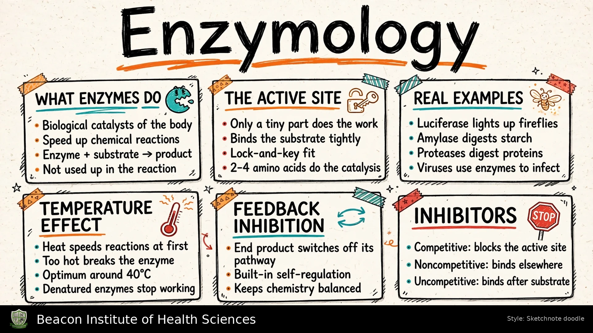
15. Reading the accompanying Enzymology infographic
The gallery illustration gives a quick visual introduction. Its compact labels need the detail provided in these notes. In the complete catalytic cycle, enzyme plus substrate forms enzyme plus product; the enzyme is regenerated. "Too hot breaks the enzyme" usually refers to disruption of the functional protein structure, not necessarily cleavage of its peptide chain. "Optimum around 40 degrees Celsius" must not be treated as a universal enzyme optimum. The temperature response depends on the particular enzyme and assay conditions.
The number of amino-acid residues that directly participate in catalysis varies; "2-4 amino acids" is an illustrative simplification rather than a rule for all enzymes. A small catalytic region is supported by a larger protein structure that positions it correctly. Lock-and-key is an introductory model, while induced fit explains the dynamic binding changes more fully. Inhibition should be interpreted using the precise kinetic distinctions in Section 9, including the difference between mixed and pure noncompetitive inhibition.
The examples show the range of enzyme functions. Amylase hydrolyzes starch, proteases hydrolyze proteins and luciferase catalyzes a light-producing reaction. Some viruses encode enzymes such as polymerases or proteases that support replication and processing, while also relying on host-cell machinery. These examples illustrate biological roles; they do not imply that all enzymes have identical structures, cofactors or temperature preferences.