
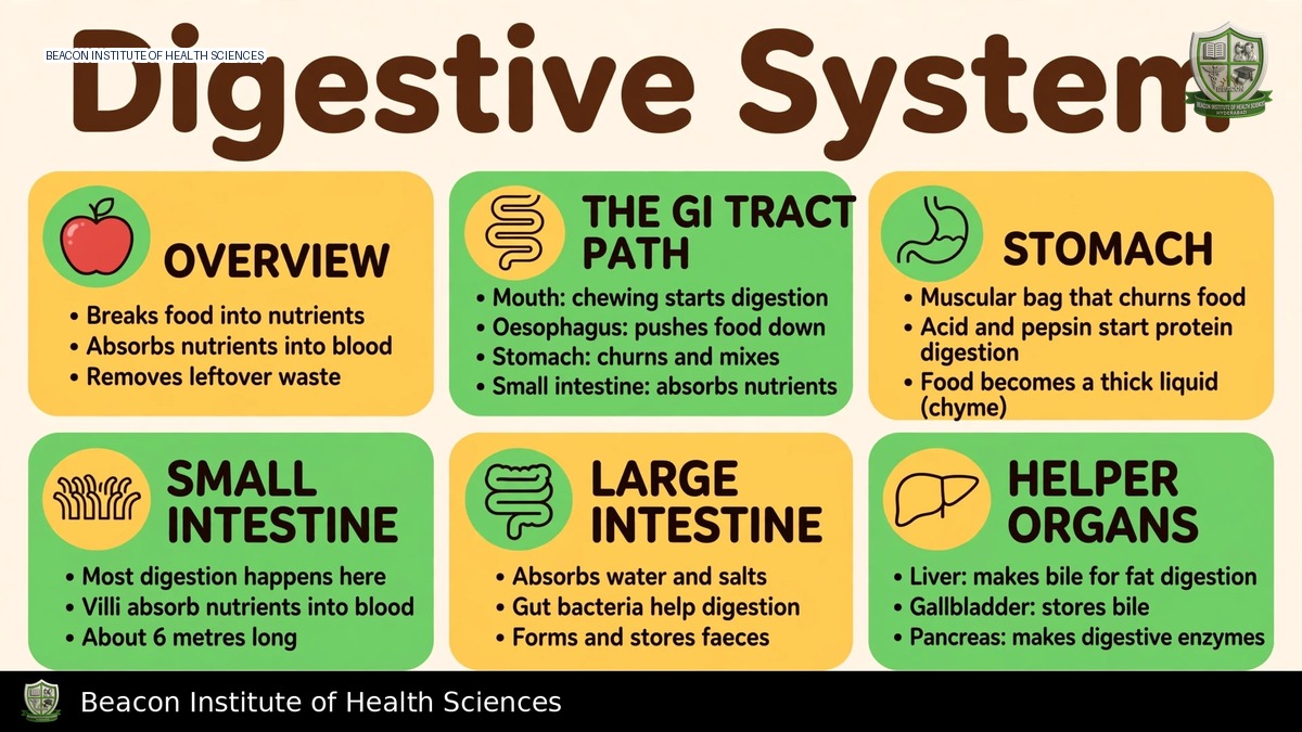
Learning Objectives and Functions
This Anatomy and Physiology I unit establishes normal digestive structure and function, rather than focusing on dietary planning or the management of gastrointestinal disease. The learning objectives include tracing the alimentary canal, identifying accessory organs, describing gastrointestinal wall layers, explaining motility and secretion, and distinguishing digestion from absorption and elimination.
Digestion involves ingestion, propulsion, mechanical breakdown, chemical digestion, absorption, and defaecation. Mechanical actions reduce particle size and mix contents, while enzymes break chemical bonds so nutrients can be absorbed. Absorption transfers substances from the lumen into the blood or lymph. Defaecation eliminates residual material and differs from metabolic waste excretion by organs such as the kidneys.
Canal and Accessory Organs
The digestive route encompasses the mouth, pharynx, oesophagus, stomach, small intestine, large intestine, rectum, and anal canal. Accessory organs, such as the teeth and tongue, aid in food processing. Salivary glands, the liver, gallbladder, and pancreas provide secretions or support digestion, though food does not normally pass through them. Sphincters coordinate the movement of contents between regions. The lumen is continuous with the external environment, requiring substances to cross epithelial barriers before becoming available to internal tissues.
Wall Layers and Control
From the lumen outward, the typical gastrointestinal wall comprises four layers: mucosa, submucosa, muscularis externa, and an outer serosa or adventitia, depending on the location.
Wall Layers
- Mucosa: Includes the epithelium, lamina propria, and muscularis mucosae.
- Submucosa: Contains vessels, lymphatics, and nerves.
- Muscularis Externa: Typically has inner circular and outer longitudinal layers. The stomach features an additional oblique layer.
- Serosa: Covers many intraperitoneal surfaces.
- Adventitia: Anchors other regions.
Control Mechanisms
The enteric nervous system coordinates local activity through submucosal and myenteric networks, with autonomic modulation. Peristalsis propels contents through coordinated contraction, whereas segmentation mainly mixes them. Hormones and local signals link secretion and motility to the contents. Nervous and endocrine regulation cooperate rather than functioning as wholly separate systems.
Mouth, Swallowing, and Oesophagus
Mastication and saliva moisten and break down food. Salivary amylase initiates starch digestion, while saliva also provides lubrication and oral protection. The tongue forms and moves a bolus. Swallowing involves a voluntary oral phase, followed by subsequent reflex-coordinated phases that transfer material through the pharynx and oesophagus while protecting the airway. Oesophageal peristalsis assists propulsion, and the lower oesophageal sphincter function limits reflux. Swallowing safety relies on coordinated activity, not merely gravity.
Stomach
The stomach stores food, mixes it into chyme, and begins substantial protein digestion. Gastric regions include the cardia, fundus, body, and pyloric part. Parietal cells secrete hydrochloric acid and intrinsic factor, while chief cells release pepsinogen. Acid supports the conversion of pepsinogen to active pepsin and provides antimicrobial effects, with mucus and bicarbonate protecting the surface. Intrinsic factor is crucial for vitamin B12 absorption later in the terminal ileum. The stomach absorbs selected substances but is not the primary site of nutrient absorption. Emptying is regulated by the properties of chyme and duodenal responses.
Small Intestine and Absorption
The duodenum receives gastric contents along with bile and pancreatic secretions, while the jejunum and ileum continue digestion and absorption. Circular folds, villi, and microvilli expand the surface area at different structural levels. Brush-border enzymes complete important digestive steps, and pancreatic enzymes help digest carbohydrates, proteins, and lipids. Bicarbonate helps neutralize acid.
Monosaccharides and amino acids predominantly pass into the capillary blood and portal circulation. Many long-chain lipid products are assembled into chylomicrons and enter lacteals before venous return. Water absorption accompanies solute transport. The terminal ileum is important for the absorption of intrinsic-factor-associated B12 and bile salts. Different nutrients utilize different mechanisms and regions; it is important to avoid assigning all absorption to one short segment.
Liver, Gallbladder, and Pancreas
The liver produces bile, processes portal-delivered nutrients, stores selected substances, synthesizes many plasma proteins, and participates in metabolism and detoxification. Bile salts support emulsification and micelle formation; bile is not an enzyme that directly hydrolyzes dietary fat. The gallbladder stores and concentrates bile, releasing it through biliary pathways when stimulated.
The pancreas contains exocrine acini and ducts that supply digestive enzymes and bicarbonate to the duodenum, and endocrine islets that secrete hormones like insulin and glucagon into the blood, representing distinct functions. Secretin promotes bicarbonate-rich secretion in response to acid, while cholecystokinin supports pancreatic enzyme secretion and gallbladder contraction in response to appropriate nutrients. Gastrin supports gastric secretory activity. Feedback mechanisms also limit secretion and movement when downstream processing requires it.
Large Intestine and Defaecation
The large intestine includes the caecum, appendix, ascending, transverse, descending, and sigmoid colon, rectum, and anal canal. It absorbs remaining water and electrolytes, supports microbial activity, compacts material, and stores faeces. Most nutrient absorption has already occurred in the small intestine. Microbes contribute to fermentation and selected vitamin production but do not provide every nutrient required by the body.
Rectal distension initiates reflex activity. The internal anal sphincter is composed of smooth muscle, while the external anal sphincter is skeletal muscle with voluntary control. Defaecation integrates autonomic, spinal, and voluntary influences. Continence depends on sensation, pelvic support, motility, and functional circumstances, not solely on one sphincter.
Nursing Application and Revision
Nurses should relate anatomy to clinical assessments such as swallowing assessment, abdominal observations, hydration status, bowel-pattern history, and nutritional status within their clinical scope. It is crucial to document symptoms precisely and escalate concerning pain, persistent vomiting, bleeding, or swallowing difficulty according to local procedures. Nurses must not infer a treatment or diagnosis from a simplified organ diagram alone.
For revision, students should practice tracing the path of food, pancreatic secretion, bile, and portal blood. Match stomach cell types to their products, compare villi with microvilli, and distinguish the liver's bile production from gallbladder storage. Explain how a carbohydrate product reaches portal blood and how a typical long-chain dietary lipid reaches lymph. Recognize that normal physiology supports, but does not replace, clinical assessment and patient-specific plans.