
1. Core Concepts & Definitions of Elimination
Metabolic processes continuously produce by-products and waste substances from ingested food and fluids that must be expelled to preserve physiological homeostasis. The biological mechanism responsible for this clearance is elimination.
A. Definition of Elimination
Elimination is the physiological process of excreting waste products and undigested substances formed within the body, transporting them from the internal environment to the outside of the body. This encompasses both the excretion of fluid/water by-products through the urinary tract (urinary elimination) and solid food residues through the gastrointestinal tract (fecal elimination).
B. The Elimination Pattern (Gordon's Functional Health Patterns)
In professional nursing practice, the Elimination Pattern is assessed as a core component of Marjory Gordon's Functional Health Patterns. It describes the regulation, control, and removal of bodily by-products and wastes, specifically evaluating the movement and expulsion of feces and urine outside the body.
When assessing an individual's elimination pattern, the nurse evaluates specific clinical parameters:
- Voiding Frequency: The number of times an individual urinates throughout a 24-hour period (e.g., whether normal voiding occurs 4 to 5 times daily or 6 to 7 times daily).
- Fecal Regularity: The timing, consistency, and frequency of bowel evacuations.
- Established Elimination Habits: The individual's behavioral routines and the physiological degree of control they maintain over their bladder and bowel functions.
2. Functional Anatomy & Pathway of the Urinary Tract
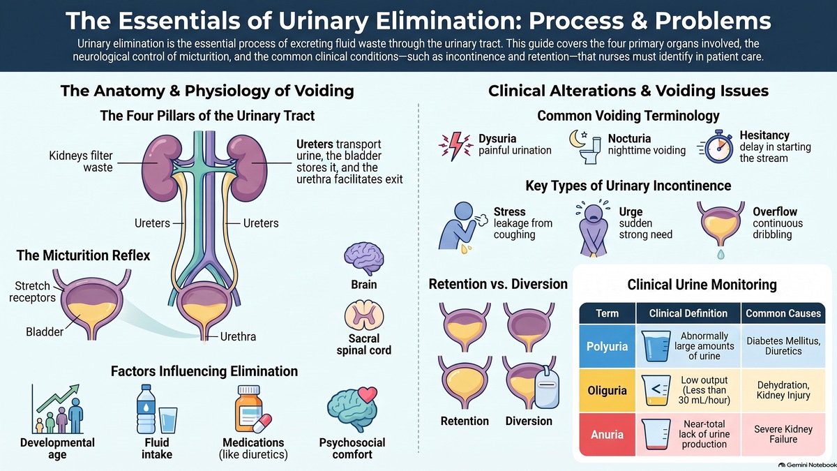
Urinary Elimination is executed by a specialized group of continuous organs that filter circulating blood, remove fluid waste products, synthesize urine, and discharge it outside the body. Together, these organs constitute the Urinary Tract.
Fluid wastes follow a precise sequential pathway:
Kidneys → Bilateral Ureters → Urinary Bladder → Urethra → External Environment (Urethral Meatus).
The Four Primary Organs of the Urinary Tract:
| Organ | Anatomical Nature | Primary Physiological Function |
|---|---|---|
| Kidneys | Primary filtration organs | Act as biological filters that remove metabolic wastes from circulating blood to form urine, simultaneously regulating fluid and electrolyte balance. |
| Ureters | Bilateral muscular tubes | Actively transport the newly formed urine from the renal pelvis of each kidney down into the urinary bladder. |
| Urinary Bladder | Hollow muscular reservoir | Serves as a temporary storage organ that holds and reserves urine until sufficient volume accumulates to stimulate the urge to void. |
| Urethra | Terminal excretory tube | Passageway through which urine travels from the bladder to exit the body externally via the urethral meatus. |
3. Clinical Terminology & the Mechanism of Urination
Standardized clinical terminology is employed to describe the normal and involuntary aspects of bladder voiding:
- Urination / Micturition / Voiding: The active physiological process of emptying the urinary bladder by discharging urine through the urethra.
- Enuresis: The involuntary passing or leakage of urine. This is typically observed in young infants and toddlers whose nervous system and urethral sphincters have not yet developed voluntary control.
- Nocturnal Enuresis (Bed-Wetting): Involuntary urination occurring during sleep at night in a child who has already attained an age where daytime voluntary bladder control has been established.
The Act of Urination: Neurological Reflex & Cortical Control
Voiding is a coordinated neuro-muscular reflex governed by the interaction between the bladder wall, the sacral spinal cord, and higher cerebral brain centers:
- Bladder Filling & Wall Distension: As urine accumulates, bladder volume increases, stretching the detrusor muscle wall.
- Sensory Afferent Activation: Stretch receptors within the bladder wall detect tension and send sensory nerve impulses to the sacral segment of the spinal cord.
- Ascending Spinal Transmission: The sacral spinal cord relays these sensory messages up to higher cortical centers in the brain.
- Cerebral Appraisal: The brain cognitively evaluates whether the physical and social setting is appropriate to initiate urination or whether the urge should be voluntarily inhibited.
- Motor Efferent Stimulation: When the decision to void is made, the brain transmits motor impulses instructing the external urethral sphincter to relax.
- Micturition Execution: Relaxation of the external sphincter, paired with detrusor contraction, allows urine to pass through the urethra and discharge through the urethral meatus.
4. Factors Influencing & Altering Voiding Patterns
Urinary elimination is highly sensitive to developmental, environmental, psychological, pharmacological, and pathological influences. Nurses must evaluate these seven primary factors during clinical assessment:
A. Developmental Factors
Infants consume an exclusively liquid diet (breast milk or formula). Because of their high fluid intake and immature neurological system (lacking voluntary sphincter control), infants void frequently, and the vast majority of metabolic wastes are excreted as urine.
B. Psychosocial Factors
Urination requires psychological comfort and privacy. Requesting a patient to collect a urine specimen in an unfamiliar clinical container often produces anxiety and hesitation. Likewise, bedridden patients who cannot walk to the toilet and must use a bedpan or bedside urinal often experience acute embarrassment and lack of privacy, which can cause temporary voiding inhibition.
C. Fluid and Food Intake
The volume and concentration of urine directly reflect fluid intake. High fluid intake expands intravascular volume, stimulating renal filtration and increasing urination. Conversely, diminished fluid consumption—such as during religious fasting (e.g., Ramadan) or severe fluid restriction—causes dehydration and drastically decreases urine formation.
D. Medications
Certain pharmacologic agents directly alter renal hemodynamics. A prime clinical example is Lasix (Furosemide), a potent loop diuretic. Diuretics inhibit electrolyte reabsorption in the renal tubules, promoting rapid fluid excretion and significantly increasing urine output.
E. Muscle Tone
Effective bladder continence requires adequate muscle tone in the detrusor muscle and urethral sphincters. If these muscles become flaccid or weak, voluntary urinary control is compromised, leading to continuous dribbling, leakage, or incomplete emptying.
F. Pathological Conditions
Medical diseases alter urinary elimination through cognitive or neurological mechanisms:
- Alzheimer's Disease: Progressive cognitive decline leads patients to forget to use the commode or lose awareness of bladder fullness, weakening the conscious urge to void.
- Neurogenic Bladder: Neurological trauma or neuropathy impairs the transmission of sensory signals from the bladder to the sacral spinal cord and brain, abolishing normal voiding urges and voluntary control.
G. Surgical and Diagnostic Procedures
Surgical interventions involving the pelvic cavity, urinary tract, or surrounding structures—as well as invasive diagnostic procedures (such as cystoscopy)—can cause direct tissue edema, mechanical trauma, or temporary reflex suppression of renal and bladder function.
5. Alterations in Urine Production & Urinary Volume
Changes in urine volume reflect alterations in renal perfusion, glomerular filtration rate, or hormonal regulation:
| Condition | Clinical Definition | Etiological Factors & Clinical Manifestations |
|---|---|---|
| Polyuria | Abnormally large volume of urine produced and excreted by the kidneys. |
|
| Oliguria | Significantly diminished urine production resulting in an output of less than 30 mL per hour. | A critical warning sign of renal compromise. Observed in:
|
| Anuria | Near-complete absence or severe cessation of urine production by the kidneys. | Indicates critical end-stage renal failure, complete bilateral urinary tract obstruction, or severe circulatory collapse requiring emergency medical intervention. |
6. Common Clinical Problems of Urinary Elimination
Disruptions in urinary elimination manifest with distinct patterns of frequency, discomfort, and retention:
| Clinical Problem | Diagnostic Features | Pathophysiological Mechanism |
|---|---|---|
| Urinary Frequency | Voiding at frequent intervals without a corresponding rise in total daily urine volume. | Daily voiding increases from the normal 3-4 times up to 7-8, 10, or more times daily, typically caused by bladder wall inflammation, infection, or irritation. |
| Nocturia | Waking from sleep 2 to 3 or more times per night to urinate. | Associated with excessive evening fluid consumption, congestive heart failure, nocturnal diuretic action, or prostatic hypertrophy. |
| Urgency | An intense, sudden, and uncontrollable urge to void. | The sensation is so overwhelming that patients must rush to the commode, occasionally experiencing involuntary leakage before reaching the toilet. |
| Dysuria | Painful, burning, or difficult urination. | Commonly caused by mucosal inflammation, bladder trauma, acute urinary tract infection (UTI), or the passage of urinary calculi (stones). |
| Urinary Hesitancy | Delay or difficulty in initiating the urinary stream. | The patient strains to initiate voiding because the muscles of the external urethral sphincter fail to relax promptly. |
| Urinary Retention | Inability to empty the bladder, leading to progressive urine accumulation. | Urine continuously pools, causing progressive bladder distension. Stagnant urine in chronic retention is a primary predisposing factor for Urinary Tract Infections (UTIs). |
7. Surgical Urinary Diversion
When normal urinary outflow through the bladder and urethra is blocked or destroyed by severe trauma, bladder carcinoma, strictures, or extensive calculus disease, surgical redirection of urine is necessary. This is known as a Urinary Diversion.
The Urostomy Procedure
Surgeons disconnect the ureters from the diseased or traumatized bladder and attach them to an isolated segment of intestine (ileal conduit). This conduit is brought through the anterior abdominal wall to create a surgically visible stoma. An external appliance (collection pouch) is placed securely over the stoma to continuously collect draining urine, which is periodically drained and discarded by the patient or nursing staff.
8. Urinary Incontinence & Its Clinical Classifications
Urinary Incontinence is the involuntary loss, leakage, or continuous dribbling of urine caused by the breakdown of voluntary sphincter control. It is classified into four major clinical categories:
| Incontinence Type | Triggering Factors & Mechanism | Clinical Presentation |
|---|---|---|
| Stress Incontinence | Occurs when an abrupt increase in intra-abdominal pressure overcomes urethral resistance. Triggered by coughing, sneezing, laughing, lifting, or physical exertion. | Sudden downward intra-abdominal pressure pushes against the bladder, forcing the urethral sphincter open and causing involuntary leaking or dribbling of urine. |
| Urge Incontinence | Triggered by hyperactive, involuntary detrusor muscle contractions creating an urgent, sudden need to void. | The urge is so abrupt and intense that the patient cannot hold, suppress, or stop micturition, resulting in involuntary voiding. |
| Mixed Incontinence | A combination of both Stress and Urge Incontinence mechanisms. | The patient experiences involuntary leakage during physical exertion/coughing as well as sudden, uncontrollable urges. |
| Overflow Incontinence | Results from chronic urinary retention where the bladder reaches its maximum anatomical distension. | Urine continuously dribbles drop by drop as new urine enters, while a large volume of residual urine remains permanently trapped in the distended bladder. |
This study guide covers the foundational principles, terminology, organs, mechanisms, etiological factors, and classifications of altered urinary elimination. Detailed nursing interventions, urinary catheterization protocols, and comprehensive nursing management of altered elimination patterns are explored in subsequent lectures within this course unit.