
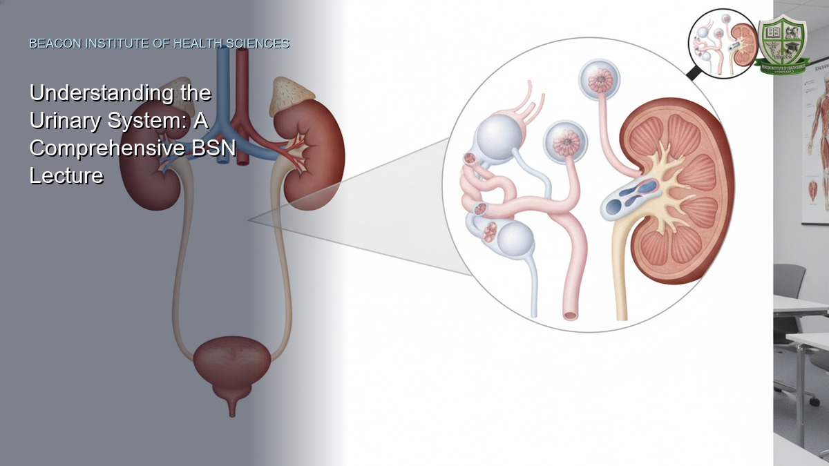
Introduction to the Urinary System
The urinary system is a vital homeostatic organ system primarily responsible for filtering blood, maintaining fluid and electrolyte balance, and eliminating metabolic wastes. Understanding its intricate anatomy and physiology is fundamental for BSN students, as it underpins various aspects of patient care, including fluid management, renal disease, and medication administration.
Functions of the Kidneys
The kidneys perform several crucial functions:
- Blood Filtration: They filter approximately 180 liters of blood plasma daily, removing metabolic wastes, toxins, and excess ions.
- Water Processing: They regulate water volume and ensure proper hydration or elimination of excess water.
- Waste Elimination: Kidneys are the primary organs for eliminating nitrogenous wastes (urea, uric acid, creatinine) and various drugs from the body.
- Blood Pressure Regulation: They help regulate blood pressure by influencing blood volume and producing regulatory substances.
- Acid-Base Balance: Kidneys maintain the correct balance between acids and bases in the body fluids.
- Hormone Production: They stimulate the production of erythropoietin, a hormone essential for red blood cell production in the bone marrow.
- Vitamin D Activation: Kidney cells convert vitamin D to its active form, crucial for calcium absorption and bone health.
Organs of the Urinary System
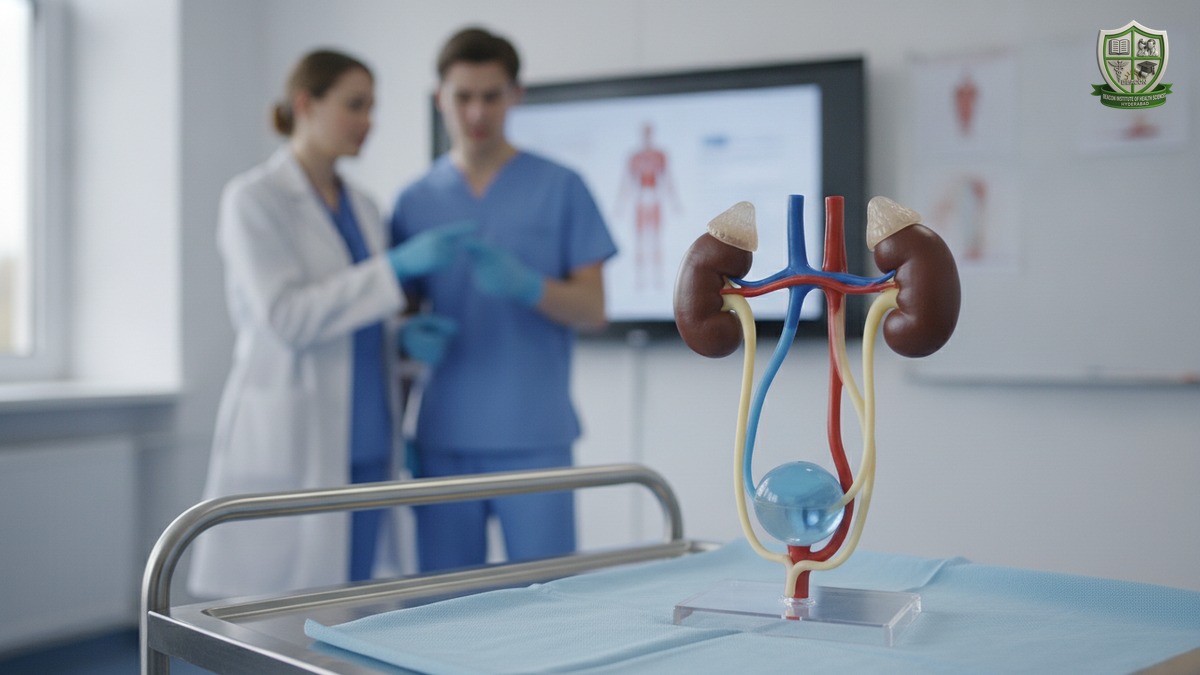
The urinary system comprises four main organs:
- Two Kidneys
- Two Ureters
- One Urinary Bladder
- One Urethra
Kidneys: Location and Anatomy
The kidneys are paired, bean-shaped organs located in the upper abdominal cavity. They are retroperitoneal, meaning they lie behind the peritoneum, extending from the T12 to the L3 vertebrae. The right kidney is typically slightly lower than the left due to the liver's presence.
External Anatomy
- Size: Approximately 5 inches (12.5 cm) long, 2.5 inches (6 cm) wide, and 1 inch (3 cm) thick.
- Hilum: A medial indentation where the renal artery enters, and the renal vein and ureter exit.
- Adrenal Gland: Sits atop each kidney, functioning as a separate endocrine organ.
Kidney Coverings
The kidneys are protected by three layers:
- Fibrous Capsule: A transparent, fibrous membrane that tightly encloses the kidney, providing protection and maintaining its shape.
- Adipose Capsule: A fatty mass surrounding the fibrous capsule, cushioning the kidney against physical trauma.
- Renal Fascia: The outermost layer, anchoring the kidney to surrounding structures and the posterior abdominal wall.
Internal (Gross) Anatomy
A sagittal view of the kidney reveals distinct regions:
- Renal Cortex: The outer, light-colored region.
- Renal Medulla: The deeper, darker, reddish-brown region, located deep to the cortex.
- Renal Pyramids: Triangular regions within the medulla, characterized by a striped appearance, with their broad bases facing the cortex and their tips (papillae) pointing towards the renal pelvis.
- Renal Columns: Extensions of cortical tissue that separate the renal pyramids.
- Renal Pelvis: A flat, funnel-shaped tube continuous with the ureter, found in the kidney's medial region. It collects urine.
- Calyces (Minor and Major): Cup-shaped areas that enclose the renal papillae, collecting urine from the pyramids and funneling it into the renal pelvis.
Microscopic Anatomy: The Nephron
The nephron is the functional unit of the kidney, responsible for forming urine. Each kidney contains over a million nephrons. A nephron consists of two main parts:
- Renal Corpuscle:
- Glomerulus: A knot of specialized capillaries where blood filtration occurs.
- Bowman's Capsule (Glomerular Capsule): A cup-shaped structure that surrounds the glomerulus and collects the filtered fluid (filtrate).
- Renal Tubule: A long, twisted tube extending from Bowman's capsule. It includes:
- Proximal Convoluted Tubule (PCT): Extends from Bowman's capsule.
- Loop of Henle: A hairpin-shaped loop with descending and ascending limbs.
- Distal Convoluted Tubule (DCT): Follows the loop of Henle.
- Collecting Duct: Receives urine from multiple DCTs and carries it through the medulla to the renal pelvis.
There are two main types of nephrons:
- Cortical Nephrons: The most common type (85%), located almost entirely within the renal cortex, with a short loop of Henle.
- Juxtamedullary Nephrons: Have long loops of Henle that extend deep into the renal medulla, playing a crucial role in concentrating urine.
Blood Supply to the Kidneys
Blood enters the kidney via the renal artery, which divides into progressively smaller arteries: segmental, interlobar, arcuate, and cortical radiate (interlobular) arteries. These lead to afferent arterioles, which feed the glomeruli. Blood leaves the glomerulus via the efferent arteriole, which then forms a network of peritubular capillaries surrounding the renal tubule, essential for reabsorption and secretion. Blood then drains into venules and eventually into the renal vein.
Ureters: Structure and Function
The ureters are slender tubes, about 25-30 cm long, that transport urine from the renal pelvis of the kidneys to the urinary bladder. They primarily function through peristaltic contractions of their muscular walls. Their superior ends are continuous with the renal pelvis, and their inferior ends pass through the abdominal cavity, behind the peritoneum, before entering the posterior wall of the bladder.
Ureters have three layers:
- Outer Fibrous Layer: Connective tissue.
- Middle Muscular Layer: Smooth muscle for peristalsis.
- Inner Epithelial Layer: Transitional epithelium, allowing for stretch.
Urinary Bladder: Structure and Function
The urinary bladder is a smooth, collapsible, muscular sac that temporarily stores urine. Its location is in the pelvic cavity, posterior to the pubic symphysis.
- Function: Temporary storage of urine.
- Wall Composition: Composed of smooth muscle (detrusor muscle) and lined with transitional epithelium, enabling significant stretching without damage.
- Trigone: A triangular region on the bladder base outlined by the openings of the two ureters and the urethra. This area is clinically significant as infections tend to persist here.
Urethra: Structure and Function
The urethra is a thin-walled tube that carries urine from the bladder out of the body by peristalsis. It contains two sphincters:
- Internal Urethral Sphincter: Involuntary smooth muscle, located at the bladder-urethra junction, preventing urine leakage between voiding.
- External Urethral Sphincter: Voluntary skeletal muscle, located where the urethra passes through the pelvic floor, allowing conscious control over urination.
The length and function differ between sexes:
- Female Urethra: Approximately 3-4 cm long, opening anterior to the vaginal opening.
- Male Urethra: Approximately 14-20 cm long, extending through the penis and serving both the urinary and reproductive systems (carrying urine and semen).
Urine Formation: Three Steps
Urine formation involves three distinct processes within the nephron:
- Glomerular Filtration:
- Movement of substances from the blood into the renal tubule.
- Water and small solutes (e.g., glucose, amino acids, ions, urea) are forced from the glomerulus into Bowman's capsule.
- Proteins and blood cells are too large to pass through the filtration membrane.
- Tubular Reabsorption:
- Movement of needed substances from the filtrate back into the blood.
- As filtrate passes through the renal tubule, essential substances like water, glucose, amino acids, and some ions are reabsorbed into the peritubular capillaries.
- Tubular Secretion:
- Movement of unwanted substances from the blood into the filtrate.
- Hydrogen ions, potassium ions, creatinine, and certain drugs are secreted from the peritubular capillaries into the renal tubule, further eliminating them from the body.
Characteristics of Urine
Normal urine exhibits specific characteristics:
- Volume: Average adult urine output is 1.0 to 1.8 liters per 24 hours, but can range from 0.8 to 2.0 liters depending on fluid intake and other factors.
- Components: Contains nitrogenous wastes (urea, uric acid, creatinine), sodium ions, potassium ions, phosphate, and sulfate ions.
- Color: Generally clear and ranges from pale yellow to deep yellow, depending on hydration and diet.
- Odor: Typically sterile and slightly aromatic when fresh.
- pH: Averages around 6.0 but can vary significantly (4.5 to 8.0) based on diet and metabolic activity.
- Specific Gravity: Usually ranges from 1.002 to 1.035, indicating the concentration of solutes. Pure water has a specific gravity of 1.000.
Micturition (Urination)
Micturition is the process of emptying the urinary bladder. It is initiated by:
- Bladder Filling: As urine accumulates, the bladder wall stretches.
- Activation of Stretch Receptors: When the bladder volume reaches about 200-400 ml, stretch receptors in the bladder wall are activated.
- Neural Reflex: These receptors send impulses to the spinal cord, triggering a reflex arc.
- Detrusor Contraction: Parasympathetic nerve impulses cause the detrusor muscle to contract.
- Sphincter Relaxation: The internal urethral sphincter relaxes involuntarily.
- Voluntary Control: The external urethral sphincter, under voluntary control, can be relaxed to allow urine to flow out of the body or kept contracted to postpone urination.

Conclusion
The urinary system, with its complex network of organs and sophisticated mechanisms, is indispensable for maintaining the body's internal environment. A thorough understanding of kidney functions, the anatomy of the urinary tract, the intricate process of urine formation, and the mechanics of micturition is critical for BSN students. This knowledge forms the foundation for effective nursing assessment, intervention, and patient education in various clinical settings.