
Core Terminology of Mobility and Body Mechanics
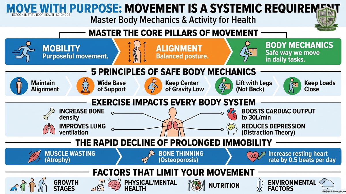
Understanding the mechanics and movement of the human body is fundamental to nursing practice. Nurses must master core terminology to assess, plan, and execute care for patients facing mobility limitations or requiring structural rehabilitation. The following four concepts form the core foundation of this unit:
- Mobility: The ability of an individual to move freely, easily, rhythmically, and purposefully within their environment. Purposeful and coordinated movement is a critical indicator of functional independence, muscular strength, and neurological integration.
- Joint Mobility: The degree of movement (range of motion) that can occur at an articulation—the structural point where two bones meet (an anatomical joint). True joint mobility represents unrestricted movement before being limited by surrounding soft tissues, such as muscles, tendons, ligaments, and cartilage.
- Body Alignment and Posture: The geometric and spatial relationship among the head, shoulders, spine, hips, knees, and ankles when the body is in any position (sitting, standing, or lying down). Proper body alignment aligns these structures along a vertical line, minimizing strain on the musculoskeletal system, reducing pressures on the vertebral spine, and preventing muscle fatigue.
- Body Mechanics: The coordinated and safe utilization of the human body during movement, posture maintenance, and daily living. It encompasses how we hold, position, and balance our body weight while standing, walking, lifting, carrying, bending, pushing, and pulling. Proper body mechanics protect both the patient and the nurse from musculoskeletal injury.
The Principles of Safe Body Mechanics and Lifting
Nursing care is physically demanding and involves frequent lifting, moving, and repositioning of clients. To perform these duties safely and prevent debilitating occupational injuries (especially lower back strain), nurses must apply established physical principles of body mechanics in every clinical action:
- Proper Alignment: Always maintain proper body alignment when lifting, carrying, or moving objects. This distributes forces evenly across skeletal structures and avoids placing concentrated stress on the lower back.
- Wide Base of Support: A wide base of support increases overall stability. The base of support is defined as the area occupied by a person's feet when standing. Spreading the feet slightly apart (shoulder-width) expands this base, lowering the risk of losing balance.
- Low Center of Gravity: Keeping the center of gravity low maximizes stability. In humans, the center of gravity shifts with posture, and a lower posture (e.g., flexing the hips and knees) increases steadiness.
- Equilibrium and Gravity Alignment: An object's equilibrium is maintained as long as its line of gravity passes directly through its base of support. If the body shifts so that the line of gravity falls outside the base of support, balance is lost and fall risk increases.
- Leverage and Muscle Selection: The stronger the muscle group used for a task, the greater the amount of work they can safely perform. Nurses must utilize large, powerful muscle groups (such as the quadriceps, gluteals, and hamstrings of the legs) rather than smaller, weaker muscles (such as those in the lower back).
Step-by-Step Clinical Guidelines for Safe Lifting:
Nurses must strictly follow these mechanical steps to protect themselves and patients during lifting operations:
- Plan the Lift: Assess the weight of the object or client. Determine if mechanical assistance or additional staff members are required. Do not attempt a lift alone if there is any doubt about safety.
- Widen Your Base of Support: Position your feet firmly on the ground, shoulder-width apart, to establish a stable foundation.
- Bend Your Knees: Squat down toward the load by flexing your knees and hips. Never bend forward at the waist or keep your knees locked, as this forces the spinal column to bear the entire load.
- Tighten Abdominal Muscles: Contract your stomach and core muscles. This acts as an internal support corset, stabilizing the pelvis and lumbar spine during exertion.
- Lift with Your Leg Muscles: Push upward using the powerful muscle groups of your thighs and buttocks. Let your legs do the work while keeping your spine stable.
- Keep the Load Close to Your Body: Hold the weight close to your torso. This minimizes the lever arm and the torque placed on your lower back, dramatically reducing the effort required.
- Keep Your Back Straight: Maintain a neutral spinal curve throughout the lift. Avoid twisting your spine while carrying or lifting; instead, pivot with your feet to change direction.
The Principles of Gravity and Balance
Every physical movement occurs within a constant gravitational field. To understand patient balance, postural stability, and the mechanics of locomotion, nurses must analyze three central physical principles:
- Center of Gravity: The specific point in an object or human body where the gravitational force is concentrated, and where weight is equally distributed on all sides (also referred to as the center of mass). When standing erect, the human center of gravity is located in the pelvis. As long as this point is stabilized, the body remains in a state of physical equilibrium and will not fall.
- Line of Gravity: An imaginary, vertical line that runs directly through the center of gravity, dividing the body's mass into two equal halves. In a balanced standing posture, the line of gravity extends vertically from the top of the head, passes through the ear, down through the trunk and pelvis, and drops to the ground exactly between the feet. Maintaining body balance requires that muscles constantly adjust to keep this line centered.
- Base of Support: The physical area of contact that supports the body's weight. The closer the center of gravity is to the ground, the more stable the base of support becomes. Spreading the feet wider apart provides a broader base of support, significantly enhancing physical steadiness. This principle is utilized by weightlifters and athletes who expand their foot positioning to handle heavy loads without tipping.
Factors Affecting Patient Mobility
A patient's ability to move and exercise is not solely determined by physical strength. It is a complex outcome influenced by developmental, physiological, psychological, and environmental factors:
- Growth and Development: An individual's age and musculoskeletal maturity dictate their posture, body proportions, bone density, muscle mass, motor coordination, and protective reflexes. Infants have rapid growth and primitive reflexes; toddlers undergo postural changes; adolescents experience rapid height and muscle mass development. In older adulthood, reflexes slow, bone density decreases, and joint flexibility naturally declines, affecting overall mobility.
- Physical Health: Any physical disease, trauma, or congenital defect can severely impair movement. For example, cardiorespiratory illnesses limit the oxygenation and nutrient delivery necessary for sustained muscle contractions, rendering patients weak and easily fatigued.
- Mental Health: A patient's desire and motivation to move are highly dependent on mental health. Psychological disorders, particularly clinical depression or chronic stress, can deplete a patient's initiative to exercise. This is often visually evident in a 'slumped' posture, with a lowered head, rounded shoulders, and an overall lack of physical energy or drive.
- Nutrition: Nutritional status directly impacts physical capacity. Undernutrition leads to progressive muscle wasting, bone mineral loss, and severe generalized weakness. Conversely, overnutrition leading to obesity places immense mechanical stress on joints and bones, making movement physically demanding, painful, and highly restricted.
- Personal Values and Lifestyle: An individual's habits are heavily influenced by their background. Families that value physical fitness, sports, and outdoor activities pass these healthy behaviors down to their children. Conversely, families accustomed to a sedentary lifestyle foster inactive behaviors, which carry forward into adulthood.
- External Factors: The physical environment can facilitate or obstruct movement. Extreme ambient temperatures (excessive heat or freezing cold) and unfavorable weather conditions (such as heavy rain or snow) act as severe barriers to outdoor physical activity and exercise.
- Prescribed Limitations: In clinical settings, a patient's mobility may be intentionally restricted by medical orders. Examples include prescribed strict bed rest, immobilization via orthopedic casts, skeletal traction, surgical restrictions, or protective limitations for severe cardiac conditions where elevating the heart rate and respiratory demand would be dangerous.
Systemic Benefits of Physical Activity and Exercise
Regular physical activity and targeted exercise act as powerful therapeutic interventions, promoting optimal functioning across every major physiological and psychological system of the human body. The clinical benefits are outlined below by system:
- Musculoskeletal System: Exercise increases joint flexibility, preserves and enhances muscle strength, and maximizes joint range of motion (ROM). Crucially, weight-bearing exercise stimulates bone mineral deposition, preserving bone density. This is achieved by maintaining a healthy balance between osteoblasts (bone-forming cells) and osteoclasts (bone-resorbing cells).
- Cardiovascular System: Physical activity elevates heart rate and strengthens the myocardium, enhancing its contractile force. It optimizes blood supply to the heart muscle and peripheral tissues. During heavy exertion, exercise can increase cardiac output up to 30 liters per minute in conditioned athletes (compared to a resting normal of approximately 5 liters per minute).
- Respiratory System: Exercise increases pulmonary ventilation and the depth of respiration to meet the body's elevated oxygen demands. It optimizes gas exchange in the alveoli, improves oxygen delivery to the tissues, and strengthens respiratory muscles.
- Gastrointestinal (GI) System: Movement stimulates appetite, enhances digestive efficiency, and accelerates gastrointestinal motility. By facilitating peristalsis (the wave-like contractions of the GI tract), exercise promotes regular bowel movements and directly relieves constipation.
- Metabolic System: Regular activity boosts the basal metabolic rate (BMR). It positively alters the lipid profile by increasing High-Density Lipoproteins (HDL, 'good' cholesterol) and decreasing Low-Density Lipoproteins (LDL, 'bad' cholesterol) and total circulating cholesterol.
- Urinary System: Physical activity improves blood flow to the kidneys, optimizing the filtration and excretion of waste products. It promotes complete bladder emptying and prevents urinary stasis (the stagnation of urine in the bladder), which is a primary cause of urinary tract infections and kidney stones.
- Psychoneurological System: Exercise channels physical energy positively, generating a profound sense of well-being. It improves stress tolerance by stimulating brain cell activity, enhances cognitive function, boosts self-concept and body image, reduces clinical depression through natural biochemical changes and healthy distraction, and improves the overall quality of sleep.
- Immune System: Movement enhances lymphatic circulation, which accelerates the movement of immune cells throughout the body. This strengthens the body's primary defense 'army' (white blood cells and antibodies), accelerating the destruction of invading pathogens and preventing infectious diseases.
Pathophysiology of Patient Immobility
When a patient is subjected to prolonged immobility or a bedridden state, the lack of mechanical stress and gravity-assisted physiological processes triggers a rapid, destructive cascade of multi-system decline. This pathophysiological process is detailed below:
- Musculoskeletal System: The absence of weight-bearing stress triggers disuse osteoporosis, where osteoclast activity outpaces osteoblasts, causing calcium to deplete from bones, leaving them weak and highly brittle. Without contractions, disuse atrophy occurs, causing muscles to waste and shrink. This is dramatically illustrated by historical cases of ascetic individuals who kept a limb completely immobile (e.g., raised toward the sky) until the muscle mass vanished entirely, leaving only bare bone. Furthermore, immobility leads to contractures (the permanent shortening and tightening of muscles and tendons, freezing joints in deformed positions) as well as severe joint stiffness and pain.
- Cardiovascular System: Immobility reduces cardiac reserve, causing the heart to work harder to pump blood. Resting heart rate increases by approximately 0.5 beats per minute for each day of complete bed rest, increasing the risk of tachycardia. Orthostatic hypotension occurs because peripheral vessels lose their tone; when the patient attempts to stand, blood pools in the lower extremities under gravity, causing a sudden drop in blood pressure, cerebral ischemia, and fainting (syncope). Lack of muscle pumping leads to venous vasodilation and stasis, where blood pools stagnantly in deep veins, leaking into tissues to cause dependent edema in the lower limbs and hands. Critically, low blood flow and blood pooling promote thrombus formation (clotting). If a thrombus breaks free from a vessel wall, it becomes an embolus, which can travel through the bloodstream and block vital blood vessels in the lungs or brain—a life-threatening medical emergency.
- Respiratory System: In a recumbent position, gravity-assisted drainage is lost, causing thick respiratory secretions to pool deep within the bronchioles. This impairs ventilation and cough effectiveness, leading to atelectasis (the collapse of alveoli and lung segments, which severely reduces the surface area available for gas exchange). Furthermore, stagnant, pooled secretions provide a fertile breeding ground for microorganisms, resulting in hypostatic pneumonia—a leading cause of death in bedridden patients.
- Metabolic System: Immobility decreases the basal metabolic rate. Due to diminished energy expenditure, patients experience anorexia (loss of appetite). The body enters a state of negative nitrogen balance, where protein catabolism (breakdown) vastly exceeds anabolism (synthesis), leading to progressive muscle wasting and a high excretion of nitrogen. Additionally, negative calcium balance occurs as calcium resorbed from bones enters the bloodstream and is excreted in large amounts via urine, leaving the bones severely depleted.
- Urinary System: In a supine position, the kidneys and bladder lose the benefit of gravity for drainage, causing urinary stasis and retention. The high concentration of calcium in the urine (from bone resorption) combines with this stasis to form renal calculi (kidney stones). These stones scrape and damage the delicate lining of the urinary tract, making the mucosal membrane vulnerable to invading bacteria and leading to severe urinary tract infections (UTIs).
- Gastrointestinal System: The absence of movement, combined with a decline in abdominal muscle tone and slow peristalsis, leads to severe, obstinate constipation.
- Integumentary System (Skin): Sustained pressure on bony prominences compresses local blood vessels, cutting off the supply of oxygen and nutrients to tissues. This leads to a rapid loss of skin turgor and tissue ischemia, culminating in skin breakdown and the formation of painful, infection-prone pressure ulcers (bedsores).
- Psychological & Psychosocial Functioning: Immobility breeds an increased dependent behavior. Patients must rely on others for basic needs, leading to deep feelings of helplessness, frustration, and feeling like a burden. Over time, they may display apathy, a flat affect (becoming emotionless), extreme self-centeredness, and total social withdrawal from family, friends, and healthcare staff.
System-by-System Comparison: Exercise vs. Immobility
To help clinical nurses synthesize this vast multi-system knowledge, the table below contrasts the positive physiological outcomes of exercise with the degenerative effects of immobility:
| Body System | Therapeutic Benefits of Exercise | Pathophysiological Effects of Immobility |
|---|---|---|
| Musculoskeletal | Increases flexibility & ROM; maintains muscle strength; stimulates bone density (osteoblasts). | Disuse osteoporosis (brittle bones); disuse atrophy (muscle wasting); contractures; joint stiffness/pain. |
| Cardiovascular | Strengthens heart muscle; increases cardiac output (up to 30L/min); improves coronary flow. | Diminished cardiac reserve; resting tachycardia; orthostatic hypotension; venous stasis; dependent edema; DVT/embolism. |
| Respiratory | Elevates ventilation and depth; optimizes alveolar gas exchange; strengthens respiratory muscles. | Pooling of thick respiratory secretions; atelectasis (lung collapse); hypostatic pneumonia. |
| Metabolic | Elevates basal metabolic rate (BMR); increases HDL ('good' cholesterol); lowers LDL ('bad' cholesterol). | Decreased BMR; anorexia; negative nitrogen balance (protein catabolism); negative calcium balance. |
| Urinary | Optimizes renal blood flow; ensures complete bladder emptying; prevents waste stagnation. | Urinary stasis and retention; renal calculi (kidney stones); severe urinary tract infections (UTIs). |
| Gastrointestinal | Stimulates appetite; increases peristalsis; improves digestion; prevents constipation. | Slowed peristalsis; decreased abdominal tone; obstinate constipation. |
| Integumentary | Promotes healthy peripheral circulation and optimal skin turgor. | Local ischemia; rapid loss of skin turgor; skin breakdown; pressure ulcers (bedsores). |
| Psychosocial | Generates well-being; increases stress tolerance; reduces depression; improves sleep quality. | Dependent behavior; helplessness; frustration; apathy; flat affect; self-centeredness; social withdrawal. |
Anatomy and Physiology Review of the Musculoskeletal System
To understand normal and abnormal movement, a solid review of the anatomy and physiology of the musculoskeletal system is necessary. The system is composed of the following structural elements:
- Bones: The structural foundation of the body. The skeleton is divided into two main parts: the Axial Skeleton (including the skull, vertebral column, and the thoracic cage consisting of the ribs and sternum) and the Appendicular Skeleton (including the shoulder girdle [clavicle and scapula], the bones of the upper limbs [humerus, radius, ulna, carpals, metacarpals, and phalanges], the hip bones, and the bones of the lower limbs [femur, patella, tibia, fibula, tarsals, metatarsals, and phalanges]).
- Muscles: Contractile tissues that generate force. Major muscle groups include facial and neck muscles, trunk muscles, muscles of the upper and lower limbs, and the pelvic floor muscles.
- Joints: The junctions where two or more bones meet. Joints are classified based on mobility into: Fibrous Joints (immovable, such as skull sutures), Cartilaginous Joints (slightly movable, such as the intervertebral discs of the spine), and Synovial Joints (freely movable, containing synovial fluid and cartilage. Synovial joints include hinge joints like the elbow or knee, and ball-and-socket joints like the shoulder or hip).
- Tendons: Dense, fibrous bands of connective tissue that attach muscles firmly to bones, transmitting the force of muscular contraction to initiate movement.
- Ligaments: Tough, fibrous bands of connective tissue that connect bone to bone at joint articulations, providing joint stability and preventing abnormal, damaging movements.
Essential Functions of the Musculoskeletal System:
The musculoskeletal system serves several vital physiological functions:
- Structural Framework: Provides the overall shape, framework, and alignment of the human body.
- Muscle Attachment: Provides secure attachment points for muscles, acting as anchors for movement.
- Locomotion and Movement: Bones and muscles act together as a single functional unit to hold and move individual body parts or the entire body under voluntary control.
- Protective Boundaries: Forms rigid protective enclosures for delicate internal structures. For example, the skull protects the brain, and the bony thoracic cage forms boundaries that safeguard the heart and lungs (note that the abdominal cavity lacks bone boundaries, relying on muscles instead).
- Hematopoiesis: The bone marrow (especially red marrow) serves as the primary site for the formation of all blood cells (red blood cells, white blood cells, and platelets).
- Mineral Storage: Acts as a primary reservoir for storing vital minerals, particularly calcium and phosphorus, releasing them into the bloodstream as needed to maintain homeostasis.
- Joint Mobility: Joints act as the functional units that provide mobility and range of motion, allowing the skeleton to bend and articulate.
Characteristics of Normal Human Movement
Normal, healthy physical movement is a complex, coordinated event. It is characterized by the seamless integration of four fundamental physiological properties:
- Proper Body Alignment and Posture: Aligns body parts vertically along a central line, balancing body weight perfectly. This minimizes strain on the joints, muscles, tendons, and ligaments, prevents muscle fatigue, and ensures that internal abdominal and thoracic organs are fully supported and protected in any posture (sitting, standing, or lying down).
- Intact Joint Mobility: Requires joints to have their full, functional range of motion. Joints act as the essential pivot points of the musculoskeletal system. When muscles contract, they pull on tendons, which in turn pull on bones to execute smooth joint movements.
- Balanced Equilibrium: Maintained through a highly complex, continuous sensory and motor feedback loop. The body constantly detects its spatial position, sends feedback, and makes micro-adjustments. This balance is coordinated by communication among: the inner ear fluid (the vestibular system, located deep inside the head beneath the brain), the eyes (visual feedback), and the proprioceptors in muscles, joints, and tendons that sense tension and position.
- Coordinated Movement: Refers to smooth, balanced, and purposeful movement. It is achieved through the harmonious integration and proper functioning of three major brain structures: the cerebral cortex (initiates voluntary movements), the cerebellum (coordinates balance, posture, and fine motor control), and the basal ganglia (controls and smooths out motor activities).
The Nursing Process in Patient Mobility Management
To manage clients with actual or potential mobility limitations, nurses must apply the systematic, five-step Nursing Process (Assessment, Diagnosis, Planning, Implementation, and Evaluation) to deliver safe, grounded, and individualized care:
- 1. Clinical Assessment: The nurse gathers critical subjective and objective data:
- Subjective Data (Patient History): Assess the client's usual activity levels, exercise patterns, types of exercises performed, as well as the frequency and duration of these activities. Identify any developmental, physiological, or personal factors that influence their mobility.
- Objective Data (Physical Examination): Observe and document the client's body alignment, posture, and gait (manner of walking). Assess the range of motion (ROM) of major joints, noting any pain, swelling, or visual structural limitations (such as bandages, casts, or splints). Evaluate muscle mass and muscle strength to detect atrophy or weakness.
- 2. Nursing Diagnosis: Analyze assessment findings to formulate precise clinical diagnoses. For a client showing muscle weakness, the primary diagnosis is:
- Impaired Physical Mobility related to generalized muscle weakness (as evidenced by decreased muscle strength and restricted joint range of motion).
- 3. Planning and Goal Setting: Develop a client-centered care plan with clear, measurable goals:
- Establish goals to progressively increase the client's physical activity tolerance.
- Restore the client's ability to perform Activities of Daily Living (ADLs) independently.
- Avoid physical injury by preventing falls in vulnerable clients.
- Improve overall physical fitness, muscle strength, and joint flexibility.
- Avoid any systemic complications related to prolonged immobility (such as pressure ulcers, urinary stasis, kidney stones, UTIs, and obstinate constipation).
- 4. Nursing Implementation (Interventions): Execute targeted nursing interventions to achieve the planned goals:
- Positioning the Client Appropriately: Place the client in therapeutic, anatomically aligned positions in bed as prescribed to prevent contractures and skin pressure.
- Moving and Turning: Carefully move and turn the client in bed on a strict schedule to relieve tissue compression, prevent pressure ulcers, and maintain skin integrity.
- Range of Motion (ROM) Exercises: Perform regular active or passive ROM exercises to keep joint cartilage nourished, prevent joint stiffness, and maintain muscle tone.
- Preventing Complications: Implement specific strategies, such as encouraging hydration, performing deep breathing and coughing exercises, and promoting active leg movements to prevent deep vein thrombosis (DVT) and pneumonia.
- 5. Clinical Evaluation: Continuously assess the effectiveness of the nursing interventions and adjust the plan as needed:
- Monitor and document changes in the client's physical and mental status, and evaluate their level of motivation to participate in physical exercises.
- Review if the prescribed active or passive ROM exercises were completed and assess their specific therapeutic impact on joint flexibility.
- Evaluate if the client has successfully restored their capacity to perform Activities of Daily Living (ADLs) independently, or if further rehabilitation and assistance are required.
Clinical Practice Note: Patient positioning (including specific therapeutic positions such as Fowler's, Semi-Fowler's, Trendelenburg, Prone, Supine, and Lateral positions) and the safe mechanics of transferring clients from bed to chair are extensive and vital clinical skills. Due to their critical importance and procedural detail, these positioning techniques are compiled in a dedicated, subsequent lecture module to ensure thorough coverage without overwhelming the student.