
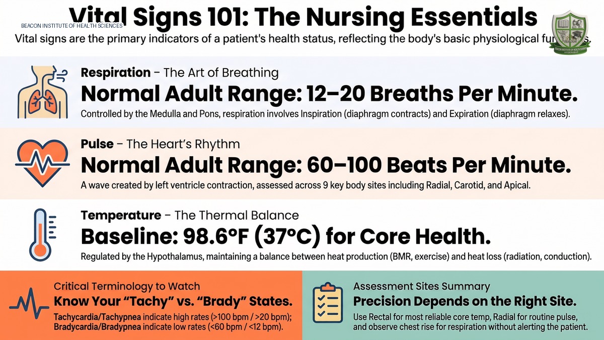
Introduction to Vital Signs in Nursing
Vital signs, also known as cardinal signs, are critical and objective measurements reflecting a patient's core physiological functions. They serve as fundamental indicators of a client's immediate health status, revealing vital systemic changes that might otherwise be unobserved. For professional nurses, accurately measuring vital signs is paramount to determine a patient's clinical state: whether they are normal, serious, or diseased. The clinical assessment of vital signs comprises six essential parameters:
- Body Temperature: The balance between heat produced and heat lost by the body.
- Pulse: The wave of blood/contraction created by the left ventricle as it pumps blood into systemic circulation.
- Respiration: The physiological act of breathing, encompassing external (gas exchange in alveoli) and internal (gas exchange in body tissues) respiration.
- Blood Pressure: The measure of pressure exerted by blood against the walls of the arteries as it flows through them.
- Oxygen Saturation (SpO2): The fraction of oxygen-saturated hemoglobin relative to the total hemoglobin in the blood.
- Pain: A highly unpleasant physical sensation caused by injury or illness, categorized into acute (short-duration, sudden) and chronic (long-term, persistent) pain.
Clinical Assessment Guidelines: When to Monitor Vital Signs
Nurses must assess and monitor vital signs at specific clinical intervals to establish baselines, track progress, and ensure patient safety. Key situations include:
- Upon Admission to a Healthcare Facility: To establish a comprehensive physiological baseline.
- Whenever a Client's Health Status Changes: This includes when a client reports new symptoms or shows signs of clinical improvement or deterioration.
- Pre-operatively and Post-operatively: Before surgery to establish a safety baseline, and after surgery to identify complications (e.g., infections leading to fever, or cardiorespiratory compromise).
- Before and After Administering Medications: Essential for evaluating the safety and efficacy of drugs that affect the cardiac or respiratory systems.
- Before and After Nursing Interventions: Required for activities that can alter cardiovascular or respiratory demand, such as patient ambulation/exercise, back care, or mouth care.
- In Intensive Care Units (ICU): High-frequency monitoring (e.g., every 30 minutes) is tracked continuously on a TPR (Temperature, Pulse, Respiration) chart to monitor acute fluctuations.
Body Temperature and Thermoregulation
Body temperature is classified anatomically into two distinct types:
- Core Body Temperature: The temperature of deep tissues, which remains relatively constant at 36.7°C to 37.0°C / 98.6°F.
- Surface Body Temperature: The temperature of skin, subcutaneous tissues, and fat, which fluctuates dynamically in response to changes in the external environmental temperature.
The Thermoregulation System
The body maintains temperature equilibrium through Thermoregulation, a negative feedback system controlled by the brain. This system consists of three primary components:
- Sensors (Thermal Receptors): Located in the skin and core tissues. The skin contains more cold receptors than warm receptors, allowing humans to perceive and respond to cold stimuli much faster.
- The Hypothalamic Integrator: The control center in the hypothalamus of the brain that maintains the core temperature set point.
- The Effector System: Physiological pathways that respond to hypothalamic commands to adjust heat production or heat loss.
Physiological Responses to Temperature Extremes
- In a Cold Environment: The hypothalamus commands effectors to decrease heat loss and increase heat production via shivering (involuntary muscle contractions generating heat), inhibition of sweating, vasoconstriction (blood vessels constricting and shifting deeper away from the skin surface), and hormonal release (epinephrine is secreted to elevate cellular metabolism).
- In a Warm Environment: The hypothalamus commands effectors to increase heat loss and decrease heat production via vasodilation (blood vessels widening and shifting superficially to release heat), sweating (promoting evaporative heat loss), and hormonal inhibition (epinephrine levels are decreased).
Mechanisms of Heat Production and Heat Loss
Heat production occurs through:
- Basal Metabolic Rate (BMR, highest in younger age groups and declines with age)
- Muscle activity (exercise/shivering)
- Thyroxine output (raises cellular metabolism)
- Epinephrine/norepinephrine and sympathetic stimulation
- Fever (pyrexia)
Heat loss occurs through four primary pathways:
- Radiation: Transfer of heat from the surface of one object to another without physical contact (e.g., a semi-dressed person in a room loses 50% of body heat via radiation).
- Conduction: Transfer of heat between molecules of different temperatures through direct physical contact.
- Convection: Transfer of heat away from the body by air currents (warmer air near skin rises, replaced by cooler, denser air).
- Evaporation: Vaporization of moisture from the skin (sweating) and respiratory tract. Insensible water/heat loss is the continuous, unnoticed loss of moisture and heat from the lungs and skin.
Clinical Classifications, Fever Patterns, and Phases
- Normothermia: Normal core temperature, averaging 36.7°C (98.6°F).
- Hyperthermia / Pyrexia (Fever): Temperature above normal. A high fever (e.g., 41.0°C / 105.8°F) is termed Hyperpyrexia. A patient with fever is febrile; a patient without is afebrile.
- Hypothermia: Core temperature dropping below normal due to excessive heat loss, inadequate heat production, or hypothalamic impairment. Signs include low vital signs, shivering, pale/waxy cold skin, decreased urinary output, and lack of muscle coordination.
Fever Patterns
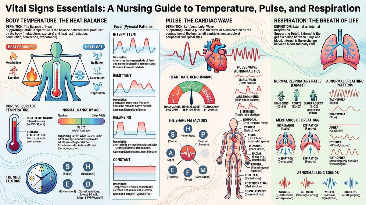
- Intermittent: Temperature alternates at regular intervals between fever and normal/subnormal ranges (e.g., Malaria).
- Remittent: Fluctuations exceed 2°C (3.6°F) in 24 hours, but temperature remains continuously above normal.
- Relapsing: Short febrile periods of a few days alternated with 1-2 days of normal temperature.
- Constant/Sustained: Temperature remains continuously elevated with minimal fluctuations (e.g., Typhoid Fever).
The Three Phases of Fever
- Onset (Chill/Cold Stage): Temperature is rising to meet new hypothalamic set point. Manifestations include shivering, pale/cold skin, piloerection ('goosebumps'), elevated heart/respiratory rates, and cyanotic nail beds.
- Course (Plateau Stage): Temperature stabilizes at an elevated peak. Shivering stops, skin becomes hot/flushed. Other signs include photosensitivity, glassy/watery eyes, severe thirst, elevated pulse/respiration, and cold sores around the mouth.
- Defervescence (Fever Abatement/Crisis Stage): Temperature falls to normal. Characterized by profuse sweating, decreased shivering, warm flushed skin, and a high risk of dehydration.
Factors Affecting Body Temperature (SHED EE)
- S - Stress: Sympathetic stimulation releases epinephrine/norepinephrine, elevating metabolism and raising temperature.
- H - Hormones: Progesterone rises during ovulation, causing a temporary temperature spike.
- E - Exercise: Muscle movement accelerates metabolism and raises temperature.
- D - Diurnal Variation (Circadian Rhythm): Natural 24-hour cycle. Temperature is highest between 8:00 PM and midnight, and lowest between 4:00 AM and 6:00 AM.
- E - Environment: Extreme external heat or cold can override thermoregulation.
- Age Variations: Newborns/neonates have immature thermoregulation, and older adults (especially above 75 years) have reduced fat, poor circulation, and lower metabolic rates, making both groups highly susceptible to hypothermia.
Anatomical Temperature Measurement Sites
| Site | Clinical Description & Advantages | Disadvantages & Risks |
|---|---|---|
| Oral (Mouth) | Highly accessible, convenient, standard for adults. | Risk of glass breakage. Seizure/child patients biting traditional mercury thermometers risk exposure to toxic mercury, causing poisoning or death. |
| Rectal (Rectum) | Most reliable/accurate site; measures core temperature directly. | Highly uncomfortable and embarrassing. Presence of stool can displace the probe, interfering with readings. Physical risk of rectal tissue injury. |
| Axillary (Armpit) | Safest, most non-invasive site with absolutely no risk of physical or organ injury. | Very slow; thermometer must be kept in place for 2 to 5 minutes to obtain an accurate reading. |
| Tympanic (Ear) | Easily accessible, comfortable, and reflects core temperature extremely fast. | Can be uncomfortable; carries physical risk of injuring or puncturing the eardrum if inserted with excessive force. |
| Temporal (Forehead) | Provides extremely rapid, non-invasive core temperature measurements. | Required equipment is highly expensive and cost-prohibitive for many healthcare settings. |
Nursing Interventions for Pyrexia (Fever)
- Monitor vital signs (temperature, pulse, respiration) and white blood cell (WBC) count (elevated WBC indicates underlying infection).
- Remove excess blankets and heavy clothing; provide warm, dry covers during the initial shivering/chill phase.
- Provide adequate nutrition and fluids (target: 2500 mL to 3000 mL of fluids per day) to prevent dehydration.
- Track and record fluid intake and output (I/O) to assess hydration and renal function.
- Provide tepid sponge baths (room-temperature water) to lower temperature through conduction and evaporation.
- Place the patient on strict bed rest to prevent muscle activity from raising metabolism and producing excess body heat.
- Administer prescribed antipyretics (acetaminophen/paracetamol, ibuprofen) to lower the hypothalamic set point.
- Administer prescribed IV therapy (Normal Saline, Dextrose Saline, Ringer's Lactate) if oral fluids are not tolerated.
- Provide routine oral hygiene to keep mucus membranes moist, preventing cold sores and secondary infections.
- Provide dry clothing and clean bed linens; prevent skin breakdown by maintaining smooth, wrinkle-free sheets.
Pulse Assessment and Hemodynamics
The pulse is a wave of blood/contraction created by the left ventricle of the heart as it pumps blood into systemic circulation. The heart is a pulsating organ, and each contraction of the left ventricle pushes blood under high pressure into the aorta, generating a wave that can be felt in peripheral arteries.
Types of Pulse
- Peripheral Pulse: Located in peripheral body parts, such as the wrist or foot.
- Apical Pulse: A central pulse located directly at the apex of the heart.
Normal Ranges of Pulse Rate Across the Lifespan
| Age Group | Average Pulse Rate (Beats per Minute) | Normal Clinical Range |
|---|---|---|
| Newborn | 130 bpm | 80 to 180 bpm |
| 1 to 3 Years | 120 bpm | 80 to 140 bpm (implied range) |
| 6 to 8 Years | 100 bpm | 75 to 115 bpm (implied range) |
| 10 Years & Teenagers | 70 bpm | 60 to 100 bpm |
| Adults & Older Adults | 80 bpm | 60 to 100 bpm |
- Bradycardia: An adult pulse rate below 60 beats per minute.
- Tachycardia: An adult pulse rate above 100 beats per minute.
Characteristics of Pulse
When assessing a patient's pulse, the nurse must evaluate five key characteristics:
- Rate: The number of beats per minute.
- Rhythm: The pattern and regularity of the beats. The time between beats must be equal and consistent.
- Volume (Stroke Volume): The amount of blood pumped with each contraction. The force/pressure felt with each beat should be equal and strong.
- Arterial Wall Elasticity: A normal healthy artery wall should feel soft, smooth, and elastic under the fingers.
- Bilateral Equality: Pulses on opposite sides of the body (e.g., right vs. left radial) should be identical in rate, rhythm, and volume.
Factors Affecting the Pulse (SHAPE FM)
- S - Stress: Sympathetic stimulation increases cardiac output and raises the pulse rate.
- H - Hemorrhage: Significant blood loss causes hypovolemia. The heart rate increases to compensate and maintain cardiac output.
- A - Age: The pulse rate is highest at birth and steadily decreases as an individual matures into adulthood.
- P - Pathology: Cardiovascular diseases and other pathologies can pathologically elevate or depress the pulse.
- P - Position Changes: Sitting or standing causes blood to pool in the lower body, reducing venous return. The heart rate increases to compensate.
- E - Exercise: Active physical movement increases metabolic demand, raising the pulse rate.
- F - Fever: Increased body temperature elevates metabolism, resulting in a rapid pulse rate.
- M - Medication: Digitalis cardiotonics decrease the heart rate, while Epinephrine (mimicking sympathetic activity) increases it.
- S - Sex (Gender): After puberty, males exhibit a slightly lower average pulse rate than females.
Anatomical Pulse Assessment Sites
| Pulse Site | Anatomical Location | Clinical Indications & Use Cases |
|---|---|---|
| Temporal | Over the temporal bone of the head, superior and lateral to the eye. | Assessed when the standard radial pulse is inaccessible. |
| Carotid | On the side of the neck, running between the trachea and the sternocleidomastoid muscle. | Used for infants, during cardiac arrest/CPR, and to evaluate brain circulation. |
| Apical | Left side of the chest, 8 cm left of the sternum, at the 4th, 5th, or 6th intercostal space. | Routine site for infants/children under 3 years, and compared with radial to identify pulse deficits. |
| Brachial | At the inner aspect of the biceps muscle, or medially in the antecubital space. | Used primarily to measure arterial blood pressure. |
| Radial | Along the radial bone, on the thumb side of the inner aspect of the wrist. | The most common, routinely used, and easily accessible site. |
| Femoral | Where the femoral artery passes alongside the inguinal ligament in the groin. | Used in cardiac arrest and to determine blood circulation to the lower extremities. |
| Popliteal | Behind the knee, where the popliteal artery passes. | Assessed to determine blood circulation to the lower leg. |
| Posterior Tibial | On the medial surface of the ankle, behind the medial malleolus. | Used to determine blood circulation to the foot. |
| Dorsalis Pedis | Along the dorsum of the foot, on an imaginary line from the ankle to the toes. | Assessed to determine blood circulation to the foot. |
Clinical Procedure for Palpating the Radial Pulse
- Prepare the Client: Inform the patient of the procedure. Assist them into a comfortable sitting or lying position. Let their arm rest alongside their body, with the palm facing downward.
- Palpate with Correct Fingers: Place 2 or 3 middle fingers flat over the radial artery. Never use your thumb because the thumb has its own strong arterial pulse, which can be mistaken for the patient's pulse.
- Count for a Full Minute: Do not count for 15 seconds and multiply by 4. Count the beats for a full 60 seconds to detect any subtle irregularities in rate, rhythm, or volume.
- Document and Report: Record the rate, rhythm, and volume accurately. Report abnormal data (such as pale or cool skin, indicating poor perfusion) to the charge nurse immediately. Never fabricate or guess entries.
Hemodynamic Pulse Contours and Abnormalities
Evaluating the shape and force of the arterial pulse wave (contour) reveals important cardiovascular details:
- Normal Pulse: Features a pulse pressure of 30 to 40 mmHg, with a smooth, rounded, and regular wave.
- Small & Weak Pulse: Characterized by diminished pulse pressure, a slow upstroke, and a prolonged peak. It is caused by a decreased stroke volume, commonly seen in Heart Failure.
- Large & Bounding Pulse: Features an increased pulse pressure, rapid rise and fall of the wave, and a brief peak. It is caused by an increased stroke volume or decreased peripheral resistance.
- Bisferiens Pulse: Characterized by a double systolic peak (two upstrokes felt per single beat). It is commonly caused by Aortic Regurgitation (where blood flows back into the left ventricle, causing a rapid double contraction) or hypertrophic cardiomyopathy.
- Pulsus Alternans: Characterized by alternating strong and weak arterial pulse amplitudes, while maintaining a regular rhythm. This is a key clinical sign of Left Ventricular Failure.
Respiratory Assessment and Pulmonary Ventilation
Respiration is defined as the physiological act of breathing. It is divided into two types:
- External Respiration: The exchange of oxygen and carbon dioxide between the alveoli of the lungs and the pulmonary capillary blood.
- Internal Respiration: The exchange of oxygen and carbon dioxide between the systemic circulating blood and the cells/tissues of the body.
The Anatomy of the Respiratory Pathway
Air enters through the nasal and oral cavities and travels through the pharynx. The epiglottis acts as a lid-like valve to prevent food from entering the airway. Air then flows down the trachea (windpipe), which is kept open by cartilage rings. The trachea bifurcates into the left and right bronchi, which enter the lung lobes and branch into smaller bronchioles, finally terminating at the alveoli (alveolar sacs) where external gas exchange occurs.
Physiology of Breathing (Pulmonary Ventilation)
Breathing is an active mechanical process divided into Inspiration and Expiration:
- Inspiration (Inhalation): The diaphragm contracts and moves downward, and the ribs move upward and outward. This causes the sternum to move forward, enlarging the thoracic cavity and lowering pressure. Air rushes in.
- Expiration (Exhalation): The diaphragm relaxes and moves upward, and the ribs move downward and inward. This causes the sternum to move inward, compressing the thoracic cavity, increasing pressure, and expelling air.
Normal Ranges of Respiratory Rate Across the Lifespan
| Age Group | Average Respiratory Rate (Breaths per Minute) | Normal Clinical Range |
|---|---|---|
| Newborn | 40 bpm | 30 to 80 bpm |
| 1 to 3 Years | 30 bpm | 20 to 40 bpm (implied range) |
| 6 to 8 Years | 19 bpm (average) | 16 to 25 bpm |
| Teenagers (16 Years) | 18 bpm | 15 to 20 bpm (implied range) |
| Adults & Older Adults | 16 bpm | 12 to 20 bpm (implied range) |
Central Control of Respiration
Respiration is controlled by the Medulla Oblongata and Pons in the brainstem. The respiratory center automatically generates and maintains the rhythm of breathing based on physiological needs. Inputs come from Chemoreceptors (which monitor drops in oxygen and spikes in carbon dioxide in the blood), Baroreceptors, the Cerebral Cortex (voluntary control), and the Hypothalamus. When CO2 levels rise or O2 drops, these receptors send impulses to the medulla and pons, stimulating the respiratory muscles to adjust breathing.
Characteristics and Terminology of Breathing
- Rate: The number of breaths per minute.
- Eupnea: Normal, quiet, effortless, and relaxed breathing.
- Bradypnea: An abnormally slow respiratory rate.
- Tachypnea / Polypnea: An abnormally fast, rapid respiratory rate.
- Apnea: The complete, temporary cessation of breathing.
- Depth: Assessed by chest expansion. Deep breathing involves a large volume of air and pronounced chest movement; shallow breathing involves a small volume of air and minimal chest movement.
- Quality: Effortless (normal) versus labored/effortful breathing (where the patient must exert physical effort and gets tired quickly).
Normal Lung Sounds (Auscultation)
Auscultating the lungs with a stethoscope reveals three types of normal lung sounds:
- Vesicular: Soft, low-pitched, rustling sounds heard over the majority of the peripheral lung fields.
- Broncho-vesicular: Intermediate intensity and pitch, heard anteriorly over the 1st and 2nd intercostal spaces, and posteriorly between the scapulae.
- Bronchial (Tracheal): Loud, high-pitched, harsh or tubular sounds heard directly over the trachea.
Abnormal Breathing Patterns
- Hyperventilation: Deep and rapid breathing that results in a large volume of air moving in and out of the lungs; commonly caused by acute anxiety.
- Hypoventilation: Shallow and slow breathing, resulting in reduced air moving into the lungs.
- Cheyne-Stokes Respiration: A highly abnormal breathing pattern where respirations start shallow, gradually increase in depth and speed, then become shallow again, followed by a period of apnea (lasting 15-20 seconds up to several minutes) before the cycle repeats.
- Dyspnea: Difficult, labored, or uncomfortable breathing, commonly referred to as Shortness of Breath (SOB). The patient feels a persistent, unsatisfied demand for air.
- Orthopnea: The inability to breathe comfortably unless sitting or standing in an upright position.
Abnormal (Adventitious) Respiratory Signs & Sounds
- Stridor: A harsh, high-pitched, crowing sound heard primarily on inspiration; it indicates a severe, life-threatening obstruction of the upper airway (larynx).
- Stertor (Snoring): A loud, snoring or sonorous sound caused by a partial obstruction of the upper respiratory tract by secretions or tissues.
- Wheeze: A continuous, musical, high-pitched whistling sound heard primarily during expiration; it is caused by air passing through partially obstructed or narrowed airways.
Factors Affecting Respiration
- Stress: Activates the fight-or-flight response, increasing the respiratory rate.
- Altitude: Higher altitudes have lower oxygen concentration, triggering an increase in respiratory rate to meet oxygen demands.
- Medications: Narcotics (such as opioids and certain analgesics) depress the respiratory center, significantly decreasing the rate and depth of respiration.
- Exercise: Accelerates metabolism and increases carbon dioxide production, raising the respiratory rate.
- Body Temperature / Environment: An increase in body temperature raises the respiratory rate (to help dissipate heat), while a decrease in body temperature lowers it to conserve heat.
Clinical Procedure for Assessing Respiration
- Determine Activity Schedule: Check the client's activity. If they have just exercised or completed a nursing intervention, wait 20 to 30 minutes for their metabolism to return to baseline.
- Maintain Client Unconsciousness: Do not inform the patient that you are counting their breaths. If they are aware, they will become self-conscious and alter their natural breathing pattern, rendering the measurement inaccurate. Pretend to take their radial pulse or write on their chart while secretly observing their chest rise and fall.
- Count and Observe: Observe one complete rise and fall of the chest as one single breath. Count for a full 60 seconds. Assess the depth (chest movement), rhythm (regularity), and quality (effortless vs. labored).
Blood Pressure Assessment and Measurement
Blood pressure (BP) is the force exerted by circulating blood against the walls of the arteries and is recorded in millimeters of mercury (mm Hg).
- Systolic blood pressure (SBP) is the upper number and reflects arterial pressure when the ventricles contract and eject blood.
- Diastolic blood pressure (DBP) is the lower number and reflects arterial pressure between heartbeats while the heart relaxes and fills.
Blood pressure is influenced mainly by cardiac output and systemic vascular resistance. Changes in blood volume, arterial stiffness, heart function, vessel diameter, medications, activity, pain, stress, and body position can therefore alter a reading.
Adult Blood Pressure Categories
| Category | Systolic (mm Hg) | Diastolic (mm Hg) |
|---|---|---|
| Normal | <120 | and <80 |
| Elevated | 120-129 | and <80 |
| Stage 1 hypertension | 130-139 | or 80-89 |
| Stage 2 hypertension | ≥140 | or ≥90 |
| Severe hypertension | >180 | and/or >120 |
These categories are for adults and follow the 2025 AHA/ACC high blood pressure guideline. Pediatric and pregnancy-related blood pressure assessment uses different criteria. A diagnosis of hypertension should not be based on a single isolated reading.
Common Factors Affecting Blood Pressure
- Age and arterial stiffness: Arterial elasticity generally decreases with age, which can raise systolic pressure.
- Exercise and activity: Temporarily increase cardiac output and can raise blood pressure.
- Stress, anxiety, and pain: Sympathetic stimulation can increase heart rate, vascular tone, and blood pressure.
- Blood volume: Fluid loss or hemorrhage can lower pressure; fluid retention can increase it.
- Medications and substances: Antihypertensives may lower BP, while some stimulants, caffeine, nicotine, and other drugs may raise it.
- Body position and technique: Unsupported posture, an incorrect cuff size, talking, or an arm positioned above/below heart level can distort the reading.
Clinical Procedure for Measuring Blood Pressure
Equipment
Use a validated upper-arm blood pressure device with an appropriately sized cuff. For manual auscultatory measurement, use a calibrated sphygmomanometer and stethoscope. An incorrect cuff size can produce inaccurate readings.
Preparation and Positioning
- Prepare the patient: Avoid caffeine, exercise, and smoking for at least 30 minutes before measurement when possible. Ask the patient to empty the bladder and rest quietly for at least 5 minutes.
- Position correctly: Seat the patient with the back supported, feet flat on the floor, legs uncrossed, and no talking. The upper arm should be bare and supported at heart level.
- Apply the cuff: Select the correct cuff size and position it snugly on the upper arm with the cuff centered over the brachial artery and the lower edge just above the elbow crease.
Manual Auscultatory Technique
- Estimate systolic pressure: Palpate the radial pulse while inflating the cuff until the pulse disappears; note this level. Inflate approximately 20-30 mm Hg above the estimated systolic level for the auscultatory reading.
- Listen over the brachial artery: Place the stethoscope over the brachial artery without pressing excessively.
- Deflate slowly: Release cuff pressure at approximately 2 mm Hg per second while listening for Korotkoff sounds.
- Identify the values: The first clear Korotkoff sound represents systolic pressure. The disappearance of Korotkoff sounds represents diastolic pressure in adults.
- Repeat when needed: For repeated measurements, allow about 1 minute between readings. At an initial assessment, measuring both arms is useful; subsequent readings are generally taken in the arm with the higher pressure when clinically appropriate.
CLINICAL ALERT: Legal Documentation Safety and Verification of Abnormal Readings
Vital signs are primary legal documents. Never fabricate, average, or guess readings. Falsified records, such as documenting normal vital signs for a deteriorating patient, represent a severe professional and criminal liability. If a parameter is abnormal, immediately re-measure, verify with a colleague or different site, implement nursing interventions, and report directly to the charge nurse.
Unexpected or very high/low blood pressure should be re-measured using correct technique and documented accurately. For adults, a reading above 180/120 mm Hg is classified as severe hypertension; if accompanied by acute symptoms such as chest pain, shortness of breath, neurologic weakness/numbness, vision change, or difficulty speaking, it requires emergency evaluation according to local clinical protocol. Never guess, average, or fabricate a vital-sign reading.
Documentation
Record the systolic and diastolic pressure in mm Hg together with clinically relevant details such as the arm used, patient position, unusual symptoms, and whether a manual or automated method was used. Recheck values that are inconsistent with the patient's condition or previous readings.