This clinical guide outlines the core nursing competencies required for skin management and the prevention of decubitus ulcers, as taught in Chapter 9 (Skin Management) of the Fundamentals of Nursing course. The target objectives covered are: defining decubitus ulcers, identifying their direct causes and risk factors, mapping common anatomical locations of sores, classifying the four clinical stages of ulceration, and applying grounded nursing interventions to maintain skin integrity and prevent tissue compromise.
Core Definition of Decubitus Ulcers
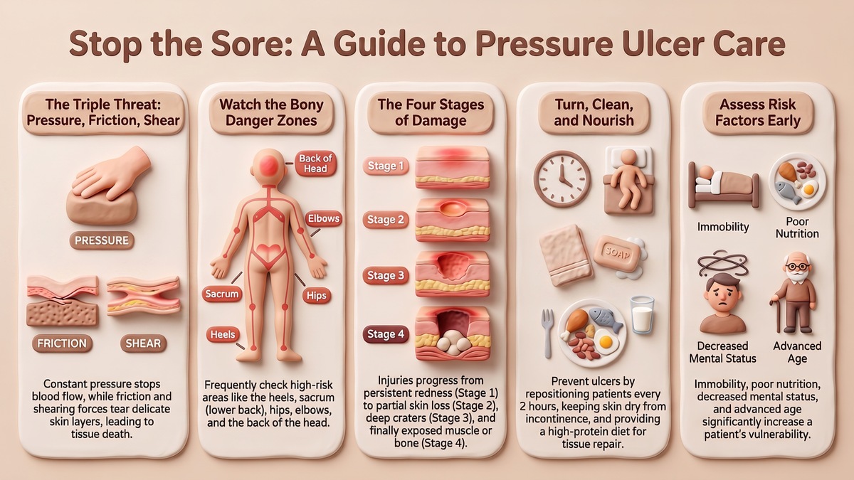
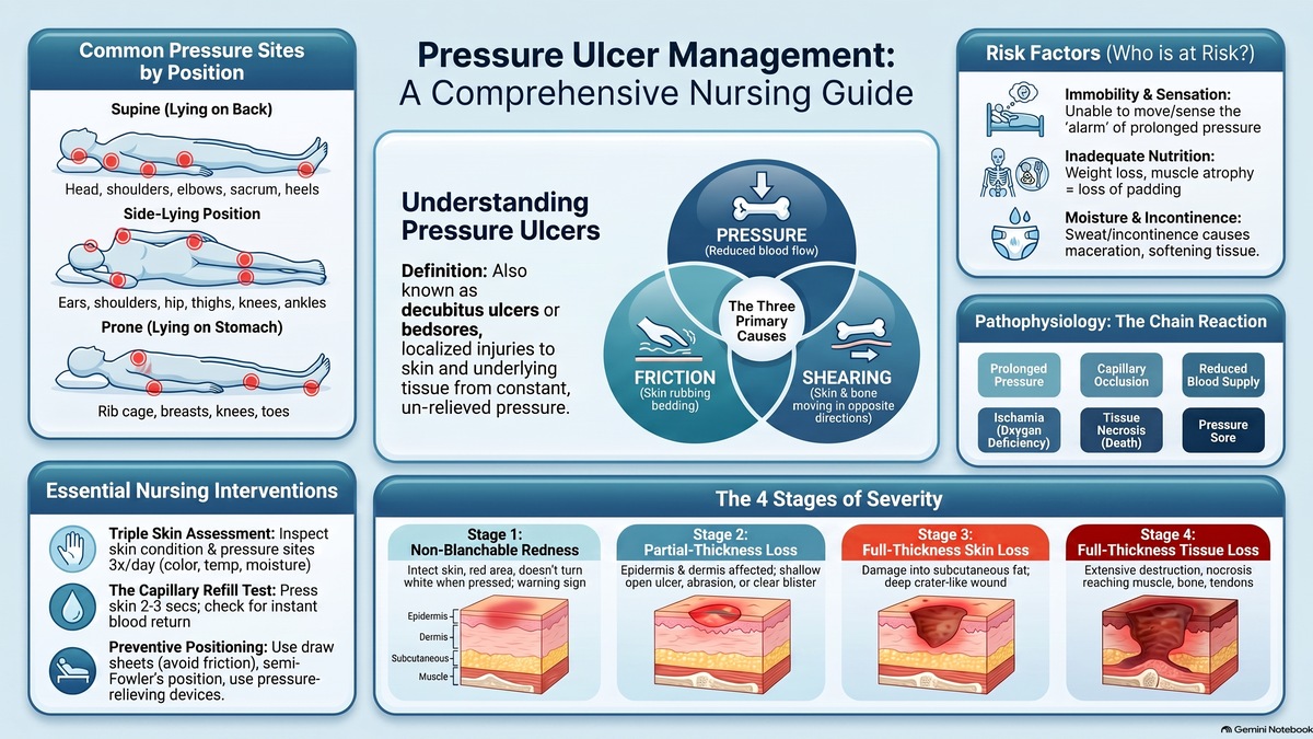
A decubitus ulcer (also known as a pressure ulcer, pressure sore, bed sore, or decubitus sore) is a localized injury to the skin and/or underlying tissue. It is caused by a constant, compressing downward force (pressure) on the body. When this constant pressure exceeds the physiological tolerance threshold that the capillary beds and tissues can withstand, it blocks local microcirculation, resulting in progressive cellular damage and necrosis of the underlying tissues.
Clinical Key: Decubitus sores develop when mechanical forces compress tissue between a bony prominence and an external surface, restricting blood flow and starving the cells of essential oxygen.
Etiology: The Three Primary Causative Factors
The clinical development of pressure ulcers is driven by three primary, overlapping mechanical and hemodynamic factors:
Constant Pressure: Constant compressing pressure applied to any localized body tissue compresses the small capillaries. This localized compression restricts or completely shuts off the blood supply (perfusion) to the tissue. Lacking blood flow, the tissues are starved of oxygen and vital nutrients, leading to tissue death (necrosis) and subsequent ulceration.
Friction: Friction occurs when the skin surface rubs directly against an external material, such as clothing, bed sheets, or blankets. Wrinkled bed sheets, improper transfer techniques, or the presence of hard debris in the bedding scrape away the protective outer epidermal layers, compromising skin barrier integrity and predisposing the site to deeper ulceration.
Shearing Forces: Shearing occurs when two adjacent tissue surfaces slide in opposite directions (e.g., when the deeper bone tissue slides downward while the superficial skin remains stationary against the bedsheets). For instance, as these opposing forces frictionally rub, they generate localized heat, which increases the oxygen demand of the cells. Since the regional capillaries are already compressed and blood flow is restricted, this combination of high metabolic demand and low supply causes rapid, severe tissue necrosis.
Pathophysiology Flowchart
Pathophysiological Stage
Physiological Mechanism & Sequelae
1. Risk Factors Act on Soft Tissue
Risk factors (such as immobility, friction, and moisture) act on soft tissue overlying a bony prominence.
2. Constant Compressing Pressure
Constant downward pressure is exerted, exceeding normal capillary pressure (occlusion of microcirculation).
3. Capillary Compression & Blockage
Capillaries and small blood vessels are folded, compressed, or completely blocked in the area.
4. Tissue Ischemia & Hypoxia
Blood supply is severely restricted, leading to localized ischemia and acute cellular oxygen deficiency (hypoxia).
5. Tissue Necrosis & Cell Death
If pressure is not immediately relieved, prolonged starvation of oxygen leads to progressive necrosis (tissue death).
6. Pressure Ulcer Formation
The necrotic tissue sloughs off, resulting in the clinical presentation of a decubitus ulcer (bed sore).
Advertisement
Clinical Risk Factors For Pressure Ulcer Development
A patient's susceptibility to developing pressure sores is influenced by multiple systemic and environmental risk factors, categorized as follows:
Immobility: Healthy individuals naturally shift position automatically when sensing pressure discomfort. However, weak, comatose, sedated, or paralyzed patients who lack movement control cannot independently change their positions. Prolonged, unrelieved compression in these patients rapidly develops into pressure ulcers.
Inadequate Nutrition: A lack of a proper, balanced diet results in rapid weight loss, muscle atrophy (muscle shortening and wasting), and the depletion of subcutaneous fat. Normally, subcutaneous fat forms a thick, cushiony pad between the bones and the skin, shielding tissues from compression. When this protective fat pad is lost, bones press directly against thin skin layers, escalating ulcer risk.
Fecal & Urinary Incontinence: Incontinence refers to the involuntary, frequent dribbling of urine or stool without the patient's conscious control. This constant presence of moisture keeps the skin persistently wet. Moisture leads to maceration—a process where wet tissue becomes extremely soft, fragile, and highly susceptible to mechanical breakdown and ulceration.
Decreased Mental Status: Patients with cognitive impairment, head injuries, or heavy sedation have a reduced mental perception of pain and discomfort. They are unable to recognize or react to prolonged pressure signals, leading to unattended tissue compression.
Decreased Sensory Sensation: Patients with sensory deficits (such as spinal cord injuries, nerve damage, or peripheral neuropathies) cannot physically feel or sense pressure or pain. Because they do not register the discomfort, they do not shift their weight, causing continuous microvascular occlusion.
Elevated Body Temperature (Fever): Fever elevates the body's Basal Metabolic Rate (BMR), which consequently increases cellular oxygen demand. If a patient is febrile, the cellular oxygen requirement skyrockets. In tissues that are already oxygen-deficient due to mechanical pressure, this hypermetabolic demand causes rapid cell death, accelerating the onset of pressure sores.
Advanced Age (Geriatrics): Aging causes a natural reduction of muscle mass and body fat (lean body), a generalized thinning of the epidermis, and vascular fragility. Older adults also experience a higher prevalence of decreased mobility and incontinence, dramatically amplifying their vulnerability to tissue breakdown.
Other Contributing Factors:
Incorrect Positioning: Subjects pressure-prone areas to friction and shearing.
Repeated Injections in the Same Area: Frequent needle pricks and localized trauma compromise skin integrity.
Hard Support Surfaces: Hard, rigid beds, stiff mattresses, or poor cushions concentrate pressure on bones.
Incorrect Application of Pressure-Relieving Devices: Improper placement of sandbags, pillows, or inflatable rings can concentrate pressure rather than relieving it.
Common Anatomical Sites of Pressure Ulcers
Pressure ulcers develop primarily over bony prominences. The specific vulnerable locations are determined directly by the patient's clinical positioning:
Patient Clinical Position
Vulnerable Bony Prominences & Pressure Sites
Supine Position (Lying on Back)
Occiput (Back of the head)
Scapulae (Shoulder blades)
Elbows
Sacrum (Lower back / Buttocks)
Heels
Lateral Position (Side-Lying)
Ears
Shoulder (Acromion process)
Elbows
Iliac crest (Hip / Greater trochanter)
Medial and Lateral Knees (where they press together)
Malleolus (Ankles / Heels)
Prone Position (Lying on Stomach)
Ears / Cheeks
Elbows
Rib cage
Breasts (especially in female patients)
Thighs
Patella (Knees)
Toes
The Four Clinical Stages of Pressure Ulcers
Pressure ulcers progress through four distinct clinical stages based on the depth of tissue destruction and damage to anatomical structures:
Stage
Pathological Feature
Clinical Presentation & Tissue Depth
Stage 1
Non-Blanchable Erythema
The skin remains completely intact (no break in the epidermis). However, there is localized redness (erythema) that is non-blanchable (it does not turn white when pressed). This stage serves as an important warning sign of impending deep tissue ulceration. The damage is superficial, confined only to the epidermal layer.
Stage 2
Partial-Thickness Skin Loss
There is a partial break in the skin involving the epidermis and extending into the dermis. This presents clinically as a superficial abrasion, a fluid-filled blister (vesicle), or a shallow, pink crater.
Stage 3
Full-Thickness Skin Loss (Subcutaneous)
Full-thickness tissue loss is present. The skin barrier is completely broken, and damage or necrosis extends through the dermis into the subcutaneous fat layer. Deep, cavernous ulceration is visible, and necrotic tissue (slough) may be present. However, the damage does not yet penetrate the underlying fascia.
Stage 4
Full-Thickness Tissue Loss (Deep)
Characterized by full-thickness tissue loss with extensive, severe destruction and tissue necrosis. The ulceration penetrates deep tissue layers, directly exposing and damaging underlying muscle, bone, tendons, or joint capsules. Necrosis is highly advanced and widespread.
Preventing decubitus ulcers requires systematic nursing assessment, monitoring, and proactive clinical care. The following interventions must be integrated into the nursing care plan:
Comprehensive Risk Assessment: Systematic assessment of all clients upon admission and periodically thereafter to identify those at high risk of skin breakdown based on their clinical status.
Frequent Skin Examinations: Thoroughly examine the patient's skin condition at least three times a day (thrice daily). Monitor skin color, temperature (excessive heat/coolness), moisture, and texture. Inspect for any localized redness or early signs of skin breakdown.
Targeted Inspection of Pressure Sites: Closely examine all vulnerable bony prominences and pressure sites corresponding to the patient's position (such as the occiput, sacrum, hips, knees, or heels).
Circulation and Capillary Refill Verification: Assess local perfusion and tissue viability by performing capillary refill checks at various skin and nail points. Press firmly on the tissue for 2 to 3 seconds, then release. If blood flow returns and the area turns pink within 2 to 3 seconds, tissue perfusion is adequate. Delayed return indicates compromised microcirculation and a high risk of tissue ischemia.
Monitoring for Incontinence: Check the patient frequently for urinary or fecal incontinence. Persistent dribbling must be identified promptly to protect the skin from prolonged moisture exposure.
Neurovascular Assessment: Regularly evaluate the patient's mental, sensory, and motor status. Assess whether the patient can feel pain or localized pressure, and ensure they are not experiencing sensory loss.
Incontinence Management Interventions: For patients experiencing incontinence, implement a structured plan:
Skin Hygiene: Immediately clean the perineal and sacral areas thoroughly after each episode.
Moisture Barrier: Apply a moisture barrier ointment over the vulnerable skin to block wetness and prevent skin maceration.
Pelvic Floor Exercises: Teach and encourage the patient to perform pelvic floor muscle exercises (Kegel exercises) to strengthen muscles and improve voluntary urinary control, significantly reducing incontinence episodes.
Minimizing Friction and Shearing Forces: Take physical precautions during transfers and positioning:
Draw Sheets: Always use a draw sheet (or friction-reducing slide sheet) to lift and slide the patient up or across the bed. Avoid dragging or pulling the patient's skin directly over the bed linens.
Slight Bed Elevation: Adjust the bed height and elevation appropriately. Avoid excessively high angles that cause the patient to slide down the bed, generating shear forces.
Bedding Management: Keep the bedsheets dry, smooth, clean, and completely free of wrinkles or debris.
Regular Position Changes: Establish a strict turning and repositioning schedule. Reposition immobile patients at least every 2 hours to relieve pressure on bony prominences.
Utilization of Comfort & Pressure-Relieving Devices: Deploy specialized supports:
Support Surfaces: Provide therapeutic mattresses, pillows, or cushions to distribute body weight evenly.
Bony Prominence Support: For comatose, paralyzed, or high-risk patients who cannot shift weight, use specialized air-filled or water-filled cushions, gloves, or balloons under bony areas (such as heels and elbows) to float and suspend these sites, eliminating direct contact pressure.
Treatment Guidelines for Developed Bed Sores
In cases where a pressure ulcer has already developed, the nursing team must follow these core clinical treatment guidelines to facilitate healing and prevent secondary infections:
Pressure Relief (Offloading): Immediately change the patient's position so that absolutely no weight or pressure is placed on the affected bony prominence. Complete offloading is essential for tissue recovery.
Wound Cleansing: Cleanse the wound bed thoroughly using prescribed solutions to keep the site clean.
Debridement: Remove dead, necrotic, or damaged tissue (slough/eschar) from the wound to promote healthy granulation and prevent bacterial colonization.
Wound Dressing: Apply sterile dressings designed to protect the tissue, manage exudate, and maintain a clean, moist, and optimal wound healing environment.
Antibiotic Therapy: Administer oral or topical antibiotics as prescribed by the healthcare provider to prevent or treat localized infections.
Nutritional Support: Provide a healthy, high-protein diet. Proper nutrition boosts the immune system, supports tissue repair, and accelerates wound healing.
Caregiver and Family Education: Educate the patient's family members and primary caregivers on proper repositioning, safe transfer techniques, and daily skin inspection to maintain care consistency outside the clinical setting.