
1. Nucleic Acids as Informational Molecules
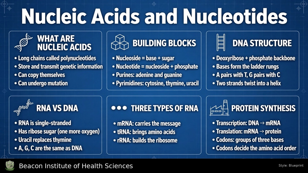
Nucleic acids are biological polymers whose subunits are nucleotides. Their two principal cellular forms are deoxyribonucleic acid, DNA, and ribonucleic acid, RNA. DNA provides a durable store of genetic information; different RNAs help express, process and regulate that information. A sequence of bases carries information, whereas the repeating sugar-phosphate framework provides the chain's structural backbone.
A polynucleotide is a chain containing many nucleotide residues. An oligonucleotide is a relatively short chain. Neither term identifies a protein: proteins consist of amino-acid residues joined by peptide bonds. In contrast, nucleic-acid residues are connected through phosphodiester linkages. Distinguishing the subunit and its linkage helps prevent confusion between these major classes of macromolecule.
For BSN study, these molecules connect biochemistry with inheritance, cell division, tissue growth, protein production and molecular testing. Their roles extend beyond a static description of the chromosome: cells must copy DNA, make appropriate RNA products and maintain nucleotide supplies for these processes.
2. The Three Components of a Nucleotide
A nucleotide contains a nitrogenous base, a five-carbon sugar and one or more phosphate groups. The sugar is ribose in a ribonucleotide and 2'-deoxyribose in a deoxyribonucleotide. The base distinguishes individual nucleotides within either family. Phosphate contributes charge and supplies the chemical connections involved in many nucleotide reactions.
- Base: a nitrogen-containing purine or pyrimidine.
- Pentose: the sugar joining the base to the phosphate-bearing portion.
- Phosphate: one or more phosphate units, commonly attached at the sugar's 5' position in free cellular nucleotides.
DNA and RNA contain related but different nucleotide sets. Changing the sugar from ribose to deoxyribose is a chemical change, not simply a change in the length of the polymer. Similarly, ATP is a particular nucleotide, rather than a name for every nucleotide or every nucleic acid.
3. Purines and Pyrimidines
Purines have two fused rings: adenine, A, and guanine, G, are the familiar purine bases of DNA and RNA. Pyrimidines have one ring: cytosine, C, thymine, T, and uracil, U, are the principal bases discussed in introductory nucleic-acid chemistry. The different arrangements of chemical groups on these rings allow specific base-pairing interactions.
The standard DNA alphabet is A, G, C and T. The standard RNA alphabet is A, G, C and U. Thus, adenine, guanine and cytosine occur in both, whereas uracil occupies the role normally assigned to thymine in RNA sequences. This description concerns the standard alphabets; biological RNA can also contain chemically modified bases.
A base alone is not a nucleotide. Adenine must be distinguished from adenosine and from adenosine monophosphate. Keeping these three names separate identifies what components are actually present in the molecule.
4. Ribose, Deoxyribose and Prime Numbering
The sugar carbons are numbered 1', 2', 3', 4' and 5', read as one prime through five prime. Prime marks distinguish sugar positions from the separately numbered atoms of the base. The base attaches to the 1' carbon. The 3' hydroxyl is important for extension of a nucleic-acid chain, and the 5' position commonly bears phosphate.
Ribose has a hydroxyl group, -OH, at the 2' carbon. In 2'-deoxyribose, that position has hydrogen instead. Both sugars retain five carbon atoms: deoxy means loss of an oxygen at the indicated position, not loss of a carbon or loss of all hydroxyl groups.
These positions explain why a sequence has a 5' end and a 3' end. They also identify where the backbone connections form. A label such as 3' describes a location on the sugar, whereas a triphosphate label counts phosphate units.
5. Nucleosides, Nucleotides and Their Names
A nucleoside contains base plus sugar, without phosphate. Adding phosphate gives a nucleotide. The base-sugar connection is a beta N-glycosidic bond: the sugar's 1' carbon connects to N9 of a purine or N1 of a pyrimidine. This is different from both a phosphate ester and a backbone phosphodiester bond.
- Adenine with ribose forms adenosine; its monophosphate is AMP.
- Guanine with ribose forms guanosine; its monophosphate is GMP.
- Cytosine with ribose forms cytidine; its monophosphate is CMP.
- Uracil with ribose forms uridine; its monophosphate is UMP.
Deoxy prefixes identify corresponding deoxyribose compounds, such as deoxyadenosine and dAMP. Thymidine conventionally refers to the deoxyribose nucleoside of thymine. Exact naming matters when reading laboratory descriptions, metabolic pathways or the names of nucleotide-related medicines.
6. Mono-, Di- and Triphosphates
AMP, ADP and ATP contain one, two and three phosphate units respectively. In ATP, the phosphate closest to ribose is alpha, the next is beta and the terminal phosphate is gamma. The first sugar-phosphate connection is a phosphate ester; the connections between successive phosphates are phosphoanhydride linkages.
Comparable families include GMP, GDP and GTP, or UMP, UDP and UTP. The prefix d identifies the deoxyribose series: dATP, dGTP, dCTP and dTTP are substrates for DNA synthesis. RNA synthesis normally uses ATP, GTP, CTP and UTP.
One nucleotide precursor does not contribute all three phosphates to the finished backbone. Polymerisation releases pyrophosphate, while the incorporated residue contributes a monophosphate unit to the chain. The activated precursor and the residue in the polymer therefore require different descriptions.
7. Phosphodiester Bonds and Chain Direction
A backbone phosphate connects the 3' oxygen of one sugar with the 5' oxygen of the next. Because the phosphate is ester-linked to two sugar groups, this connection is called a phosphodiester linkage. It produces an alternating sugar-phosphate chain with bases attached as side groups.
The chain has polarity: its two ends are chemically different. Sequences are conventionally written from 5' to 3'. Polymerases extend a growing strand by adding to its available 3' hydroxyl, so synthesis proceeds in the 5'-to-3' direction.
The order of the bases must be distinguished from the repeated backbone. A change in sequence can change biological information even when the general sugar-phosphate architecture remains the same. Hydrogen bonds between paired bases are also distinct from the covalent bonds that maintain continuity within a strand.
8. DNA Double-Helix Organisation
Cellular DNA usually consists of two complementary strands arranged as a double helix. The sugar-phosphate backbones lie toward the outside, while paired bases occupy the interior. The two strands are antiparallel: one runs 5' to 3' while the other runs 3' to 5' along the same stretch.
Adenine pairs with thymine through two hydrogen bonds, and guanine pairs with cytosine through three. These pairings place a purine opposite a pyrimidine. Base stacking also contributes to helix stability; DNA stability cannot be reduced to hydrogen-bond counts alone.
The ladder analogy identifies backbones as sides and paired bases as rungs, but the molecule is twisted rather than flat. Recognition of grooves and particular sequences allows proteins to interact with DNA. A drawing of two arrows pointing in the same direction would incorrectly represent the normal antiparallel arrangement.
9. Complementary Sequences
For a strand written 5'-ATGCCA-3', its aligned complement is 3'-TACGGT-5'. If the complementary strand must also be written in the conventional 5'-to-3' direction, its sequence becomes 5'-TGGCAT-3'. Reversing the written order is necessary because the strands run in opposite directions.
In an ordinary double-stranded DNA molecule, total adenine equals total thymine and total guanine equals total cytosine. These relationships do not require adenine to equal guanine. If adenine accounts for 30% of all bases in such a molecule, thymine also accounts for 30%; the remaining 40% comprises 20% guanine and 20% cytosine.
These percentage rules apply to the paired double-stranded molecule. They should not automatically be applied to an isolated single strand or to every RNA molecule. A sequence question must identify both the molecule and the direction in which the answer is requested.
10. DNA, Chromatin, Chromosomes and Genes
In human cells, nuclear DNA associates with proteins to form chromatin. Chromosomes are organised structures containing this DNA and associated proteins. A gene is a functional segment of DNA that contributes a product, which may be an RNA or, through RNA, a protein. A chromosome contains many genes, rather than being synonymous with one gene.
The genome describes the genetic material as a whole. Most human nucleated somatic cells normally have 46 nuclear chromosomes arranged in 23 pairs. Gametes normally contain 23. These statements describe typical complements, not every cell type or every possible chromosomal condition.
Human cells also contain mitochondrial DNA. Consequently, the statement that all human DNA occurs only in the nucleus is incomplete. Genes, chromosomes and the genome describe different levels of organisation, and their terminology should remain distinct in patient education and clinical documentation.
11. DNA Replication
Replication produces DNA from a DNA template before genetic material is distributed to daughter cells. The process is semiconservative: each new double-stranded molecule contains one parental strand and one newly synthesised strand. Complementary base pairing explains how each parental sequence can guide formation of its partner.
Unwinding exposes templates. DNA polymerases select complementary deoxyribonucleoside triphosphates and extend new strands. Both new strands are made 5' to 3', even though their templates are antiparallel. This directional requirement underlies continuous leading-strand synthesis and discontinuous lagging-strand synthesis at a replication fork.
DNA does not copy itself without molecular machinery. Replication depends on enzymes, substrates and coordinated cellular processes. The expression that DNA can copy itself refers to its templating capacity, rather than an unaided chemical action by a naked DNA molecule.
12. RNA Structure and Folding
RNA generally contains ribose and the bases A, G, C and U. Many cellular RNAs are single chains, but portions can pair with complementary regions within the same molecule. Hairpins, stems and more complex folds allow RNA to form functional three-dimensional structures.
Calling RNA single-stranded does not mean it is always a straight, unpaired thread. Its local pairing and folding are especially evident in transfer RNA and ribosomal RNA. Some RNAs also catalyse reactions, showing that RNA can function as more than a passive carrier of a message.
In protein production, RNA supplies several cooperating components. The message, the amino-acid adaptor and the ribosome's RNA components have different jobs. They should not all be described as messenger RNA or assumed to be translated into proteins themselves.
13. Messenger, Transfer and Ribosomal RNA
- Messenger RNA, mRNA: carries a sequence that ribosomes use as a template for polypeptide synthesis.
- Transfer RNA, tRNA: acts as an adaptor between an mRNA codon and an amino acid. Its anticodon pairs with the codon; its amino-acid attachment region carries the appropriate amino acid.
- Ribosomal RNA, rRNA: forms essential structural and catalytic components of ribosomes together with ribosomal proteins.
These three familiar categories cooperate rather than replacing one another. The ribosome reads the message, amino-acid-bearing tRNAs interact with it, and the ribosomal machinery supports peptide-bond formation.
Other RNA classes participate in RNA processing and regulation. Thus, three major RNA types in an introductory diagram should not be interpreted as a complete list of every RNA found in human cells.
14. Transcription
Transcription makes RNA using a DNA template. RNA polymerase and associated proteins recognise appropriate regions, begin synthesis, extend the RNA and terminate the process. RNA is synthesised 5' to 3', while the template is read in the opposite direction.
For a DNA template segment written 3'-TACGGA-5', the complementary RNA is 5'-AUGCCU-3'. The corresponding coding DNA strand is 5'-ATGCCT-3'. The RNA matches the coding strand's base order with U in place of T; it is complementary to the template strand.
Not every RNA transcript encodes a protein. Human RNA polymerases produce different RNA classes: polymerase II produces nuclear protein-coding pre-mRNAs, while other polymerases produce many structural and processing RNAs. Transcription should therefore be defined as DNA-to-RNA synthesis, rather than exclusively as production of one type of message.
15. Processing of Human Pre-mRNA
A newly made nuclear protein-coding transcript is typically processed before functioning as mature mRNA. A modified guanosine cap is added to the 5' end, and a poly-A tail is added at the processed 3' end. These features support stability and the interactions required for transport and translation.
Splicing removes introns and joins exons. Exons are retained in the mature transcript; they can include untranslated regions as well as protein-coding sequence. It is therefore too broad to define every nucleotide in an exon as part of a translated protein code.
Alternative splicing can generate different transcript forms from the same gene. Processing does not mean that the RNA sequence is translated during splicing. It prepares the RNA product, after which a suitable mature mRNA can reach cytoplasmic ribosomes.
16. Codons and the Genetic Code
A codon is a group of three consecutive bases read in an mRNA sequence during translation. Four possible bases give 64 possible triplets. In the standard genetic code, 61 codons specify amino acids and three are stop signals: UAA, UAG and UGA.
AUG specifies methionine and commonly serves as the start codon. The initiation machinery determines the correct start site and reading frame; not every AUG anywhere in a message independently starts a new protein.
The code is degenerate because several codons can specify the same amino acid. This does not mean a codon has an arbitrary meaning. Maintaining the reading frame is essential: a sequence read as AUG-CCU-GAA differs from one read starting a base later. A tRNA anticodon is complementary to a codon and is not simply another name for the codon.
17. Translation and Protein Production
Translation uses an mRNA template to assemble a polypeptide from amino acids. Initiation establishes the ribosome, the appropriate message region and initiator tRNA. Elongation repeatedly brings in suitable amino-acid-bearing tRNAs, forms peptide bonds and advances the ribosome along the message.
The A site accepts an incoming aminoacyl-tRNA; the P site holds the tRNA associated with the growing chain; the E site allows an uncharged tRNA to leave. Ribosomal RNA participates in the catalytic machinery rather than merely providing an inert platform.
At a stop codon, release factors support release of the polypeptide. The product then requires appropriate folding and may undergo further modification. Nucleotide sequence directs amino-acid order, but the polypeptide is built from amino acids, not from DNA or RNA bases converted into amino acids.
18. ATP and Other Energy-Related Nucleotides
ATP, adenosine triphosphate, contains adenine, ribose and three phosphate units. Its reactions couple energy-yielding processes to processes requiring energy, including active transport, biosynthesis and mechanical work. GTP supplies energy in other reactions, including steps of protein synthesis.
A common schematic is ATP + water → ADP + inorganic phosphate. The favourable overall reaction can be coupled to cellular work. Breaking a bond alone requires energy; net energy availability reflects the entire reaction and the relative stability of its products, rather than energy appearing solely because a bond snaps.
ATP is continually regenerated. It is an immediate metabolic carrier rather than the body's principal long-term energy store. Its adenine is a base, its adenosine portion is a nucleoside, and the complete phosphorylated molecule is a nucleotide.
19. Cyclic Nucleotides and Coenzymes
Cyclic AMP, cAMP, and cyclic GMP, cGMP, are intracellular signalling molecules. Adenylyl cyclase forms cAMP from ATP, whereas guanylyl cyclase forms cGMP from GTP. They help connect receptor activation with intracellular responses, including regulation of protein kinases and some ion channels.
Phosphodiesterases break down these cyclic signals. Cyclic AMP is therefore not merely another spelling of AMP: its phosphate forms a cyclic connection in the molecule, with different functional consequences.
Nucleotide-containing coenzymes also support metabolism. NAD+/NADH and NADP+/NADPH participate in electron-transfer reactions; FAD/FADH2 is another related carrier system. These functions show why nucleotide chemistry matters even outside DNA and RNA synthesis. A coenzyme's redox role should remain distinct from the base-sequence information carried by a nucleic acid.
20. Nucleotide Supply, Recycling and Breakdown
Cells obtain nucleotides through synthesis and recycling. De novo pathways construct nucleotide components from simpler metabolic precursors. Salvage pathways reuse bases or nucleosides released during turnover. These routes support ongoing needs for nucleic-acid synthesis and other nucleotide-dependent reactions.
Ribose-5-phosphate connects carbohydrate metabolism with nucleotide supply. Ribonucleotide precursors can be converted into deoxyribonucleotide precursors for DNA synthesis. Having a purine base available is not the same as having a complete, appropriately phosphorylated DNA precursor.
Purine breakdown in humans produces uric acid, present predominantly as urate under physiological conditions. Production and elimination both influence its concentration. Excess production, reduced removal or a combination can raise blood urate; the explanation cannot always be reduced to dietary purines alone.
21. DNA Changes and Their Consequences
A mutation is a change in DNA sequence. A substitution changes a base; insertions and deletions add or remove sequence. Effects depend on the location, extent and biological context. A change can alter a protein, affect expression or have no evident functional consequence.
In protein-coding sequence, an insertion or deletion not divisible by three can shift the reading frame. A synonymous substitution can preserve the encoded amino acid. Larger changes may involve chromosome segments or chromosome number rather than one base.
Germline changes can be passed to offspring, whereas somatic changes affect a person's body-cell lineages and are not ordinarily transmitted through reproduction. DNA repair and replication fidelity help maintain sequence integrity, but the occurrence of a variant alone does not establish a diagnosis or predict disease severity.
22. Molecular Testing and Nursing Relevance
Polymerase chain reaction, PCR, amplifies a selected DNA region. Repeated cycles separate strands, allow primers to bind and extend new strands using a polymerase. Primers provide specificity, and each cycle can increase the amount of the selected target. PCR does not automatically sequence an entire genome.
Tests directed at RNA targets can first convert RNA into complementary DNA through reverse transcription. Amplification, sequencing and measurement of gene expression answer different questions. The specimen type, target and method are therefore essential when interpreting what a laboratory request actually examines.
Genetic tests can investigate changes in genes, chromosomes or relevant proteins. A nursing explanation should distinguish identification of a variant from prediction of its effect. Consent, appropriate counselling, specimen identification and confidential handling support responsible care. Follow the requested collection instructions rather than assuming that all molecular tests use identical samples or storage conditions.
23. Purine Metabolism in Clinical Observation
A uric-acid test measures a waste product related to purine breakdown; it is not a direct measurement of the amount of DNA in a person's blood. Results can be obtained from blood or urine and must be documented with the correct specimen type, unit and laboratory reference information.
Elevated urate can be associated with gout or stones, but many people with raised blood urate do not have these complications. A laboratory value alone therefore does not establish every clinical conclusion. Cell destruction during some cancer treatments can increase purine turnover and uric-acid production, which explains why monitoring may be ordered in that setting.
For nursing documentation, record the test actually performed, relevant collection conditions and reported findings. Explain the biochemical relationship between cell turnover, purines and urate without assigning a treatment or changing medication independently. The chemistry supplies a basis for understanding the request; patient assessment and the prescribed plan determine subsequent care.